
What is Prostate MRI?
Prostate MRI is a specialized magnetic resonance imaging exam focused on the prostate gland and surrounding tissues. It uses powerful magnets and radio waves to produce detailed pictures without any ionizing radiation. Doctors primarily order a prostate MRI to detect or evaluate prostate cancer – for example, to identify suspected tumors, measure their size, and check if cancer has spread beyond the prostate. This advanced imaging can also help diagnose non-cancerous conditions like prostatitis (infection) or benign prostatic hyperplasia (BPH) by revealing inflammation or enlargement. In short, a prostate MRI provides clarity about prostate health that other tests alone (such as blood tests or ultrasound) might not achieve.
Why choose Intedia for this exam? Intedia’s radiology center in Tijuana offers state-of-the-art MRI scanning technology and a team of experienced radiologists dedicated to accurate diagnosis and patient care. Our specialists use prostate MRI to get high-resolution images that guide your treatment decisions, whether you are from Mexico or visiting from the U.S. We emphasize compassionate service alongside technical excellence, so patients feel confident that they are in good hands throughout the process.
Importance of Prostate MRI
Early and accurate detection of prostate issues is crucial for effective treatment. A prostate MRI can reveal clinically significant tumors that need treatment while helping avoid overdiagnosis of low-risk cancers. Traditional methods like a PSA blood test or a digital rectal exam (DRE) provide important clues but have limitations. PSA levels can be elevated due to non-cancerous causes (such as BPH or infections) and do not pinpoint tumor location. MRI, by contrast, gives a direct look at the prostate’s structure and any suspicious lesions. It offers more detailed images of the gland than other imaging tools like ultrasound or CT scans.
Importantly, prostate MRI has transformed the approach to biopsies. In the past, an elevated PSA often led straight to an invasive biopsy guided by transrectal ultrasound. Those “blind” systematic biopsies take multiple tissue samples at random and can miss aggressive cancer while often finding insignificant tumors. MRI changes this paradigm by identifying where significant cancer is likely or unlikely. If the MRI shows a clearly suspicious area, a targeted biopsy can be done directly at that spot for higher accuracy. If the MRI looks normal and reassuring, unnecessary biopsy can sometimes be safely deferred with close monitoring. In other words, prostate MRI helps ensure that patients get the right intervention at the right time. It’s one reason why many specialists now recommend getting an MRI before deciding on a biopsy, as high-quality evidence and guidelines in the U.S. and Europe support this MRI-first strategy. At Intedia, we prioritize such evidence-based practices – our radiologists use prostate MRI results to help your urologist determine the most appropriate next steps, potentially sparing you from an avoidable procedure.
How Prostate MRI Works
MRI uses a strong magnetic field and radiofrequency pulses to generate images of the body’s internal structures. During a prostate MRI, the machine’s magnet aligns hydrogen atoms in the body, then radio waves briefly alter that alignment. When the atoms return to their original state, they emit signals that are captured by the scanner and converted into high-resolution images. These images show cross-sectional “slices” of the prostate gland from multiple angles, allowing radiologists to examine the anatomy in detail. Because MRI relies on magnetic fields and not X-rays, it doesn’t expose you to radiation, making it a safe choice for imaging the prostate even if multiple follow-up scans are needed.
Most modern prostate MRI exams are multiparametric MRI (mpMRI) scans. This means they combine different imaging sequences to evaluate the prostate tissue in several ways. For instance, Intedia’s prostate MRI protocol includes high-resolution T2-weighted images (which show the prostate’s anatomy and any tumors), diffusion-weighted imaging (which detects how water molecules move in the tissue), and often dynamic contrast-enhanced imaging (which tracks blood flow in the gland). These advanced techniques highlight differences between healthy tissue and potential cancer. Specifically, measuring water diffusion and blood perfusion in the prostate helps identify areas that could be clinically significant cancer. Our radiologists at Intedia are experts in performing and interpreting these multiparametric studies, ensuring that no detail is missed in your prostate MRI scan.
The Technology Behind It
The clarity of a prostate MRI largely depends on the quality of the MRI machine and coils used. Intedia operates high-field MRI scanners with dedicated coils to maximize image resolution. In some cases – especially with older 1.5 Tesla magnets – an endorectal coil is used. This is a special device placed inside the rectum during the scan to bring the antenna closer to the prostate, improving signal quality. The coil is a thin latex balloon with a coil wire inside; once inserted, it is inflated to sit snugly next to the prostate and then removed after the scan. While the thought of an internal coil may sound uncomfortable, it typically causes only mild pressure and helps produce very clear images. However, many newer MRI systems (such as Intedia’s high-resolution units) can obtain excellent prostate scans without needing to use an endorectal coil, thanks to stronger magnets and better external antenna arrays.
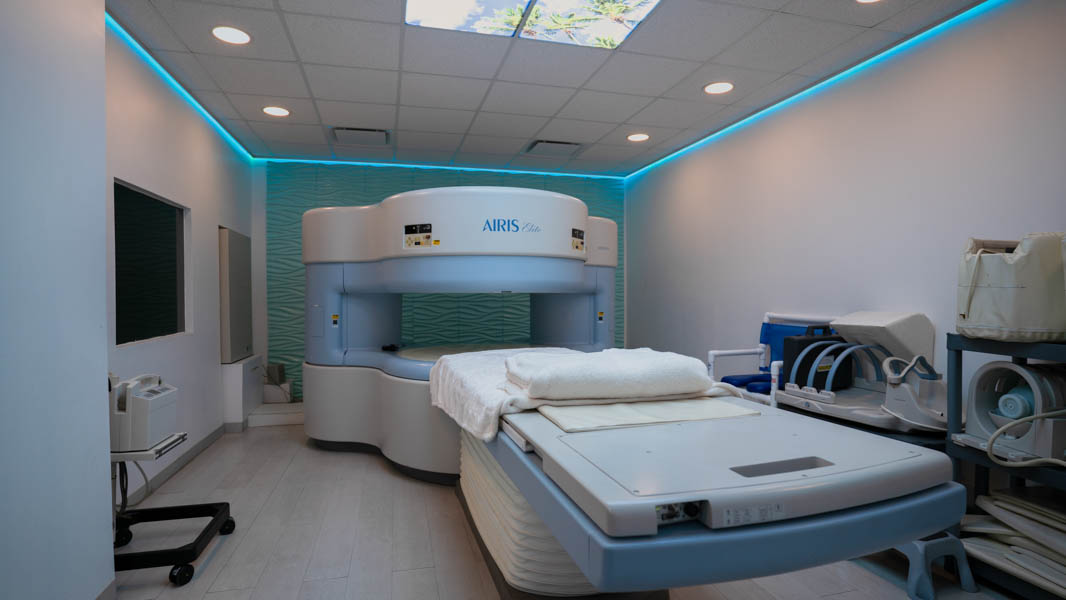
Another consideration is magnet strength. Most prostate MRIs are done on either 1.5 Tesla or 3 Tesla MRI machines – the “Tesla” rating refers to the magnet’s strength. A 3T scanner provides a higher signal and finer detail than a 1.5T, which can improve the detection of prostate lesions. Intedia’s imaging center uses cutting-edge equipment (including open MRI options) to ensure both high image quality and patient comfort. Our open MRI provides a wider, less confining space for those who are anxious or have larger body types, without significantly compromising the clarity needed for diagnostic accuracy. The combination of powerful MRI technology and specialized coils means that when you undergo a prostate MRI at Intedia, you benefit from crisp images that help doctors make confident decisions about your health.
Imaging Techniques
As mentioned, a prostate MRI exam involves multiple imaging techniques in one session. Each technique provides unique information about the prostate’s condition. The cornerstone is the T2-weighted image, which shows the prostate’s anatomy in high detail – on these images, the peripheral zone of the prostate (where many cancers arise) appears as a different shade than the central gland, and any tumors often stand out as dark or irregular areas. Another key sequence is the diffusion-weighted imaging (DWI). Areas of tumor tend to restrict the diffusion of water molecules, so they show up as bright spots on DWI and as dark areas on the corresponding ADC map (a quantitative diffusion map). This helps in identifying lesions that might not be obvious on the regular images.
Dynamic contrast enhancement (DCE) is an imaging technique where a gadolinium contrast agent is injected into a vein during the MRI. Rapidly growing tumors often have abnormal blood vessels, causing them to enhance (light up) earlier and wash out faster on serial MRI images after contrast injection. By capturing images every few seconds, radiologists can see these patterns of blood flow. A suspicious prostate lesion might show an early and intense uptake of contrast compared to normal tissue. Not every prostate MRI includes DCE; in some cases, an abbreviated exam called biparametric MRI is done, omitting the contrast. Research has shown that a faster biparametric MRI (which uses just T2 and diffusion sequences) can be nearly as effective as a full multiparametric MRI for detecting clinically significant cancer. This faster approach is gaining interest because it reduces scan time, cost, and the need for IV contrast. At Intedia, we can perform either approach based on the latest evidence and each patient’s needs. Our radiologists follow international protocols (such as PI-RADS, the Prostate Imaging Reporting and Data System) to ensure that the MRI techniques and interpretation are standardized and of highest quality. This means your prostate MRI results will be reliable and comparable to those from top institutions worldwide.
Preparing for a Prostate MRI
Proper preparation helps ensure your prostate MRI produces clear, usable images. Before the day of your scan, you will receive personalized instructions from Intedia’s staff, but general guidelines apply. You may be asked to follow a light diet the day before your MRI and to avoid ejaculation for about 2–3 days prior to the scan. Abstaining from sexual activity temporarily can prevent recent ejaculation from affecting the appearance of the prostate fluid on the MRI, which might otherwise obscure details. On the day of the exam, it’s usually recommended to eat a light meal a few hours beforehand and avoid anything that might cause gas or bloating. Some facilities request fasting for a few hours if contrast dye will be used, but in most prostate MRI cases, you can eat a small meal to stay comfortable unless instructed otherwise.
You might also be asked to do a rectal enema on the morning of the MRI or the night before. An enema empties the rectum of stool and gas, which helps reduce image distortions and improves the clarity of prostate images. Not all centers require this, but if it’s recommended, it can make a noticeable difference in image quality. Before your scan, remember to remove all metal objects (jewelry, belts, watches, etc.) since MRI’s magnet can attract metal and interfere with the scan. You will change into a gown, and a locker will be provided for your belongings. If you have any medical implants (like a pacemaker, cochlear implant, or metal clips), inform our team ahead of time. Some implants are MRI-safe, while others may pose a risk or require special arrangements. Intedia’s technologists are trained to evaluate any implant or device and will work with your referring doctor to confirm it’s safe to proceed. Overall, the preparation steps for a prostate MRI are straightforward and our team will guide you through each one to ensure you are ready for a successful scan.
Patient Preparation Tips
Even once the basic requirements (diet, fasting if needed, enema, removing metal) are handled, there are additional tips that can improve your MRI experience. Hydration is helpful – unless you’ve been told to restrict fluids, drink water before the exam. Being well-hydrated can make it easier for staff to start an IV if one is needed for contrast. If you tend to feel cold, wearing socks can keep you comfortable in the MRI room, which is often kept cool. For patients with claustrophobia (fear of enclosed spaces), proactively managing anxiety is key. One tip is to practice relaxation techniques such as deep breathing or visualization on the days leading up to the MRI. When you arrive at Intedia, let our team know if you feel nervous; we have experience helping claustrophobic patients. Sometimes something as simple as using an eye mask or a mild sedative pill (if prescribed by your doctor) can make a big difference in tolerating the scan. Intedia also offers an open MRI scanner for those who are extremely anxious – this machine has a wider opening and can significantly reduce the sense of confinement.
Plan to arrive a little early to your appointment so you’re not rushing. This gives you time to check in, use the restroom, and ask any last-minute questions. If you think you might need a sedative, arrange for someone to accompany you, because you will not be able to drive afterwards. Our staff at Intedia will walk you through a safety questionnaire regarding any allergies or medical conditions. This is the time to mention if you’ve had any prior reactions to MRI contrast dye or if you have kidney issues, since that could affect the use of gadolinium. However, the gadolinium contrast used in prostate MRI is generally very safe; allergic reactions are rare, and our team is prepared to manage them if they occur. By following these preparation tips and communicating openly with the Intedia team, you can help ensure your prostate MRI goes smoothly and comfortably.

What to Expect During the Procedure
When you undergo a prostate MRI, knowing what will happen can help you feel more at ease. After changing into a gown, you’ll be brought into the MRI suite where the scanner is located. The MRI machine is a large, tube-like structure open on both ends. You will lie flat on a cushioned table that slides into the center of the machine. For a prostate MRI, you will typically lie on your back. Our technologist will position a device called a surface coil over your pelvic area – this device receives the signals that create the images. If contrast dye is part of your exam, an IV line will be placed in your arm before you begin. Through this IV, you may receive an injection of gadolinium contrast midway through the scan. You might also receive an injection of a medication like Buscopan (or a similar antispasmodic) at the start of the procedure. This medication isn’t a sedative; it’s used to temporarily slow down bowel movements, since a moving bowel can blur the images of the nearby prostate. It can sometimes cause a dry mouth or mild blurred vision for a short while after, but these effects wear off quickly.
Once you are ready and lying comfortably, the technologist will slide the table into the MRI bore (the opening of the machine). It’s normal for the space inside the MRI to feel a bit confined – the walls of the scanner might be just a few inches from your sides. However, you’ll have earplugs or headphones on to reduce the noise, and you can still communicate with the MRI operator via an intercom. The machine will make loud thumping or knocking noises during the imaging sequences; these sounds are perfectly normal. Try to relax and hold still. The scan usually lasts around 30 to 45 minutes in total, broken up into several shorter image runs of a few minutes each. Between those sequences, the technologist may provide updates like “a couple more minutes for this next set of images.” Knowing that a caring professional is monitoring you from the control room can help you stay calm and focused on the goal: obtaining the best possible images for your health.
Risks and Benefits of Prostate MRI
Prostate MRI is a very safe procedure with numerous benefits. One major benefit is that MRI provides excellent soft tissue detail without using any harmful radiation. This makes it a preferred imaging modality for the prostate, especially when repeat scans might be needed over time (such as during active surveillance of a known cancer). The detailed images from MRI can identify aggressive tumors that need treatment and distinguish them from indolent cancers that can be left alone, which helps avoid overtreatment. MRI is also noninvasive – aside from the possible IV contrast, it doesn’t involve any surgical intervention. This stands in contrast to a biopsy, which carries risks like bleeding or infection. In fact, by incorporating MRI into the diagnostic pathway, many men can avoid an unnecessary biopsy if the scan doesn’t show suspicious findings. If a significant tumor is present, MRI is very useful in staging: it can show whether the cancer is likely confined to the prostate or if it has begun to extend through the capsule or into nearby structures, information crucial for treatment planning.
The risks associated with a prostate MRI are minimal. The procedure itself might cause discomfort for those who dislike tight spaces or loud noises, but these are managed with patient-friendly measures (open MRI, ear protection, slight sedation if needed). Gadolinium contrast, when used, has a very low incidence of allergic reactions – severe reactions are rare, and mild effects like nausea occur infrequently. In people with severe kidney dysfunction, gadolinium can pose a risk of a rare condition called NSF, so our radiologists carefully screen for kidney issues before deciding on contrast. If an endorectal coil is utilized, there is an extremely small risk of causing a tear or injury to the rectum, but this is exceedingly uncommon and coils are handled gently to avoid such issues. Overall, the diagnostic benefits of a prostate MRI – catching a serious cancer early, guiding precise treatment, and sparing men from unnecessary procedures – far outweigh the minimal risks for the vast majority of patients. Intedia’s approach further enhances safety: our strict protocols and experienced staff ensure that every precaution is taken, from double-checking your implant history to monitoring you throughout the scan.
Interpreting MRI Results
After your prostate MRI, the images will be carefully analyzed by a specialized radiologist. At Intedia, our board-certified radiologists have extensive experience in prostate imaging, which means they know how to spot even subtle abnormalities on the scans. The radiologist will look at all the sequences (T2, diffusion, contrast if given) and identify any areas that appear suspicious for cancer or other conditions. One outcome of this analysis is a structured report, which usually includes a PI-RADS score for any findings. PI-RADS (Prostate Imaging Reporting and Data System) is a standardized scoring system used internationally to describe how likely it is that a particular MRI finding is a clinically significant cancer. Scores range from 1 (very low suspicion) up to 5 (very high suspicion of aggressive cancer). For example, a small area that is probably just normal tissue or prostatitis might be labeled PI-RADS 2 (low likelihood of significant cancer), whereas a dark, irregular lesion in the peripheral zone that restricts diffusion and enhances early would likely be PI-RADS 5 (very high likelihood of cancer). By using these consistent criteria, our radiologists ensure that your urologist can understand the results clearly and make informed decisions.
The MRI report will also document other details: the size and exact location of any lesion, whether it abuts key structures like the neurovascular bundles or seminal vesicles, and if there are any signs the cancer has spread beyond the prostate. Often, measurements of the prostate gland volume are given, and any incidental findings in the pelvis (like a lymph node or a bladder finding) are noted too. Rest assured, the Impression or conclusion section of the report will highlight what, if anything, is concerning on the MRI. That section is typically the takeaway message for your urologist. If the report says something like “No MRI evidence of clinically significant prostate cancer,” that’s obviously great news. On the other hand, if a suspicious lesion is noted, the report will recommend correlation with a biopsy. Through every step, the Intedia team ensures you have access to your images and results promptly. Many patients appreciate that we can send electronic copies of their MRI scans to them or their doctors, facilitating easy second opinions or consultations as needed. Your MRI results are a vital piece of your health information, and understanding them properly – with professional guidance – will empower you to make informed decisions about your prostate health.
Discussing Results with Your Doctor
Once the prostate MRI is completed and the radiologist’s report is sent out, the next step is a discussion with your healthcare provider. This is often your urologist or the physician who ordered the MRI. In that conversation, you’ll review what (if anything) the MRI found and what it means for you. It’s a good idea to come prepared with any questions you have after reviewing the report yourself. For example, you might ask: What does a PI-RADS 4 lesion mean for my situation? Should I get a biopsy to confirm if it’s cancer? Or, if the MRI was normal, what should be the plan for future monitoring of my prostate? Your doctor will use the MRI results alongside your PSA trend, physical exam, and personal risk factors to make recommendations. If the MRI identified a concerning area, the likely next step is a targeted biopsy of that area, because imaging alone cannot provide a definitive diagnosis – tissue analysis is needed for that. If the MRI was clear of suspicious findings, your doctor may suggest continuing regular PSA checks and another MRI in the future, rather than doing an immediate biopsy.
It’s important to have a frank and open dialogue. Sometimes the MRI results can be complex, and you might feel overwhelmed – this is normal. Don’t hesitate to ask your doctor to clarify technical terms or to explain the rationale behind their advice. If any question remains unanswered, or if you feel uncertain, you can also reach out to the imaging facility. According to patient care guidelines, radiologists are often available to discuss imaging results with patients and physicians. At Intedia, we encourage collaborative care. Our radiologists frequently consult with referring doctors, and if needed, we can arrange a meeting with the patient as well to go over the images together. We want you to feel supported in understanding your prostate MRI findings. Ultimately, the MRI is a tool to help you and your healthcare team make informed decisions. Whether it leads to immediate treatment, further tests, or simply peace of mind and a plan to watch and wait, the knowledge gained from your prostate MRI can significantly shape your health strategy. And remember, choosing a provider like Intedia means you also have a partner who will help explain and navigate those results alongside you.
Comparing Prostate MRI to Other Diagnostic Tools
Prostate MRI is one part of a larger toolkit for prostate health assessment. How does it stack up against other diagnostic methods? One common tool is the PSA test, a simple blood test. PSA screening is widely used because it’s noninvasive and accessible. However, PSA is not specific to cancer – high PSA levels can come from an enlarged prostate, infections, or other conditions. By itself, PSA can lead to uncertainty: many men with elevated PSA do not have cancer, and some with aggressive cancer might have only modest PSA rises. That’s where MRI comes in. MRI can visualize the actual prostate tissue and flag areas suspicious for tumor, making it a perfect next step when PSA is elevated or other risk factors are present. Compared to a digital rectal exam, MRI is far more sensitive. A DRE (where a doctor feels the prostate through the rectum) can only detect abnormalities on the back surface of the prostate and only if they’re large enough. An MRI can detect lesions as small as a few millimeters anywhere in the gland, long before they’d be palpable.
Ultrasound imaging is another tool frequently used in urology. The standard transrectal ultrasound (TRUS) provides real-time images of the prostate and is mainly used to guide needle placement during biopsies. However, ultrasound’s ability to distinguish tumors is limited – many prostate cancers are nearly invisible on TRUS due to its lower soft tissue contrast. MRI, on the other hand, excels at soft tissue contrast, making tumors stand out more clearly. For example, MRI will often reveal lesions that ultrasound cannot see. When it comes to biopsy, modern practice often uses MRI-ultrasound fusion techniques: an MRI performed before biopsy helps identify targets, and the MRI findings are fused with ultrasound imaging during the biopsy to sample the right areas. This combined approach has a higher success rate than blind sampling. Notably, introducing MRI in the diagnostic pathway has been shown to reduce unnecessary biopsies and associated side effects while still detecting all significant cancers.
In today’s best practice, these methods are not competitors but collaborators. PSA and DRE serve as initial screening tools; MRI provides detailed imaging for clarification; ultrasound offers a convenient way to perform biopsies or monitor treatments; and biopsy confirms the diagnosis. By using each tool for what it does best, doctors can achieve a more precise and personalized evaluation of prostate health. The adoption of MRI-first strategies in prostate cancer detection is a prime example of this synergy – fewer men undergo needless invasive procedures, yet cancers that require attention are still caught in time. Intedia stays at the forefront of this integrated approach, working closely with urologists to ensure that our advanced imaging is used effectively alongside other diagnostic modalities to improve patient outcomes.
Advances in Prostate MRI Technology
Prostate MRI is a field that continues to innovate, improving accuracy and patient comfort. One of the exciting advances in recent years is the application of artificial intelligence (AI) to MRI image analysis. AI algorithms have been trained on thousands of prostate MRI scans to learn the subtle patterns of prostate cancer. In large studies, AI assistance has demonstrated the ability to detect a few percent more clinically significant cancers than expert radiologists alone, while also reducing false alarms by about half. This could translate to fewer unnecessary biopsies for patients. Essentially, AI can act as a second reader, highlighting suspicious areas on MRI that a radiologist can then review. It’s not meant to replace human experts, but to augment them, especially as prostate MRI becomes more widespread and radiologists face larger caseloads. While AI tools for prostate MRI interpretation are still being validated and aren’t yet standard in every clinic, Intedia is closely monitoring this development. We anticipate adopting proven AI support systems in the future to continue offering cutting-edge diagnostic accuracy.
Another advance is the refinement of MRI techniques themselves. As mentioned, biparametric MRI (which skips the contrast injection) has been researched extensively and is now showing promise as a faster, more accessible option without sacrificing diagnostic performance. This is particularly important in busy healthcare systems – faster scan protocols could allow more patients to be screened and diagnosed earlier. There are also ongoing improvements in MRI hardware: newer scanners have wider bores to improve patient comfort, stronger gradients for better image clarity, and noise reduction technology to make scans quieter. Intedia remains committed to leveraging these advances for our patients. We continually update our protocols in line with the latest clinical evidence and invest in new technology that has proven benefits. By choosing a center that emphasizes innovation, you ensure that your prostate health evaluation will benefit from the best that modern medical imaging has to offer.
Patient Testimonials and Experiences
At Intedia, patient satisfaction is just as important to us as clinical excellence. We take pride in the positive feedback we receive from men who have undergone prostate MRI at our facility. Many patients comment on how smoothly and professionally their appointment went – an experience that alleviated much of the stress they initially felt. For instance, some have noted that scheduling was easy and appointment times were prompt, which is crucial when you’re anxious about a health concern. Cross-border patients from the United States often appreciate our bilingual staff and the ability to coordinate care seamlessly; they felt comfortable communicating in English and valued how our team explained every step in plain language.
One common theme in testimonials is relief at how quickly results were ready. Intedia’s commitment to quick report turnaround means that in many cases, referring doctors and patients receive MRI results the same day or within 24 hours of the scan. Patients have expressed that this fast delivery of findings greatly reduced their anxiety – waiting for test results can be one of the hardest parts, after all. “I got my report and images by email that evening,” one patient recalled, noting how it allowed him to discuss next steps with his doctor sooner than expected. Others mention the personal touches that made a difference: a technologist offering a warm blanket when the room felt cold, or a reassuring voice through the intercom saying “You’re doing great, just a few minutes left.” These details reflect Intedia’s patient-centered approach. We know that undergoing an MRI, especially for something as significant as possible cancer, can be daunting. That’s why we strive to treat every patient like family – with empathy, respect, and attentive care. The positive experiences shared in patient testimonials underscore our mission to pair advanced diagnostic imaging with genuine human compassion. When you come to Intedia for your prostate MRI, you can expect to be welcomed, heard, and cared for every step of the way.
Choosing a Facility for Prostate MRI
Deciding where to get your prostate MRI can make a significant difference in your overall experience and the quality of information you obtain. Not all imaging centers are equal when it comes to this specialized exam. Here are some factors to consider and how Intedia stands out in each:
Expertise and Accreditation
Look for a facility with board-certified radiologists who have specific experience in body imaging or uroradiology. Prostate MRI interpretation is nuanced, so you want a radiologist who has read many of them. Intedia’s radiologists fit this profile – they are highly trained and stay updated on the latest prostate MRI standards. Our center is fully accredited and follows international guidelines like PI-RADS for consistent reporting.
Technology
Having modern, high-resolution MRI equipment (ideally including 3T MRI capability) is important for obtaining clear images. Also, the availability of an open MRI or wide-bore MRI can be a plus for patient comfort. Intedia is equipped with high-performance MRI systems that deliver exceptional image quality while minimizing scan time and noise. For claustrophobic patients or those with broader body types, we offer open MRI units that provide more space without sacrificing diagnostic confidence. We also utilize dedicated coils and follow optimized prostate imaging protocols to get the best results possible from our machines.
Preparation and Care
A good facility will provide thorough instructions beforehand and personalized care during the scan. At Intedia, our team takes preparation seriously – we give each patient clear guidelines (diet, fluid intake, etc.) tailored to their exam. On the day of the MRI, our technologists ensure you’re comfortable, answer all questions, and monitor you closely (including your breathing and heart rate if you’re sedated) to keep you safe. This level of attention reflects our belief that technical excellence should go hand-in-hand with compassionate patient care.
Convenience and Speed
Consider how easy it is to schedule the exam and how quickly you’ll get results. Intedia offers flexible scheduling, including accommodating cross-border patients who may be coming from Southern California. We understand that time is valuable, so we strive to provide appointments without long wait times. Additionally, our report turnaround is very rapid – often on the same day – so your referring doctor receives the information promptly. Our clinic’s location in Tijuana near the U.S. border is an advantage for Americans seeking high-quality imaging without the prolonged scheduling backlogs sometimes found elsewhere. We even coordinate our schedule to fit yours, recognizing that travel plans and personal schedules need to be considered.
Cost and Accessibility
Prostate MRI can vary in price. Intedia is proud to offer world-class imaging at a cost often more accessible than comparable services in the U.S. We accept various payment options and work with patients to ensure they can access the diagnostics they need without undue financial burden. For international patients, the value can be especially compelling – many have found that they receive the same quality (or better) imaging for a fraction of the cost by visiting our center in Tijuana, even after factoring in travel. We are also conveniently located with two sites in the city and ample parking, making the experience as hassle-free as possible.
Intedia excels in all these areas, making it a trusted provider for prostate MRI. As our team often says: when you schedule your MRI at Intedia, you are not just getting a test – you are gaining a partner dedicated to your health.