
Understanding Sonograms
In the context of sonogram vs ultrasound terminology, it’s important to define what a sonogram actually is. A sonogram is the image produced by an ultrasound examination – essentially the visual result of the scanning procedure. It is not a separate procedure, but the product of the ultrasound. For example, many parents recall the thrill of seeing their baby on an ultrasound monitor and then receiving the printed sonogram (the black-and-white image of the fetus in the uterus) as a keepsake. In medical practice, the term sonography refers to the use of an ultrasound machine to create sonograms, and the trained technologist performing the scan is called a sonographer. These sonogram images provide a visual record of internal structures that doctors analyze for diagnostic purposes. Experts often explain it with an analogy: the ultrasound machine acts like a camera, while the sonogram is the photograph it produces. In short, a sonogram is the picture obtained from an ultrasound procedure, capturing the information that the sound waves have gathered inside the body.
Understanding Ultrasounds
By comparison, an ultrasound (also known as diagnostic sonography) is the imaging procedure itself – the process of using high-frequency sound waves to visualize internal body structures. In a discussion of sonogram vs ultrasound, it’s clear that they are two sides of the same coin. The ultrasound exam is what the medical professional actually performs, and the sonogram is the result of that exam. Ultrasound waves are higher in frequency than what humans can hear, but they can create real-time images of the body’s tissues and organs by echoing off structures. Notably, unlike X-rays or CT scans, ultrasound does not use ionizing radiation, making it a safer imaging modality for a wide range of patients (including pregnant women). Many people use the terms “ultrasound” and “sonogram” interchangeably – you might hear someone say they are going to “get a sonogram” when the actual procedure is an ultrasound. This casual mix-up can cause confusion, but medically speaking the distinction is simple: the ultrasound is the test, and the sonogram is the image. In practical terms, there is no question of one being better than the other; an ultrasound and a sonogram are part of the same process. Thus, the phrase sonogram vs ultrasound isn’t about choosing one over the other, but understanding that when your doctor orders an ultrasound, the images (sonograms) are what will be produced for analysis. Both terms ultimately refer to a single, safe and invaluable diagnostic test.
Technology Behind Sonograms
To understand the science of sonogram vs ultrasound, it helps to see how an ultrasound machine creates those images. A sonogram is produced by directing sound energy into the body and capturing the echoes. During an ultrasound exam, the sonographer uses a handheld device called a transducer (or probe) which emits pulses of high-frequency sound waves into the area of interest. These sound waves travel through the body and bounce off internal structures; each tissue reflects sound differently. The returning echoes are essentially a waveform of information – their timing, strength, and frequency shifts carry clues about the depth and density of the tissues. The ultrasound machine’s computer analyzes these echo waveforms in real time and converts them into a visual image (the sonogram) on the monitor. In simpler terms, the machine “listens” for the echoes and constructs a picture from them, very much like a sonar device mapping an underwater landscape. The image appears as various shades of gray, white, and black: tissues of different densities reflect sound with varying intensity. For instance, fluid-filled areas (like a cyst or the amniotic fluid around a fetus) let sound waves through easily and thus appear black on the sonogram, whereas solid dense structures (such as bone or a calcification) reflect strongly and show up as bright white. Softer tissues produce intermediate echoes and appear in shades of gray. This grayscale mapping allows physicians to differentiate organs and abnormalities. Sonography technology has become quite sophisticated – modern systems can capture moving images and even Doppler data. The key takeaway is that the sonogram image is an instantaneous map of how sound waves interact with the body’s internal structures. This technology behind sonograms provides a noninvasive way to visualize organs and tissues, turning invisible sound echoes into a clear picture that doctors can examine.
Technology Behind Ultrasounds
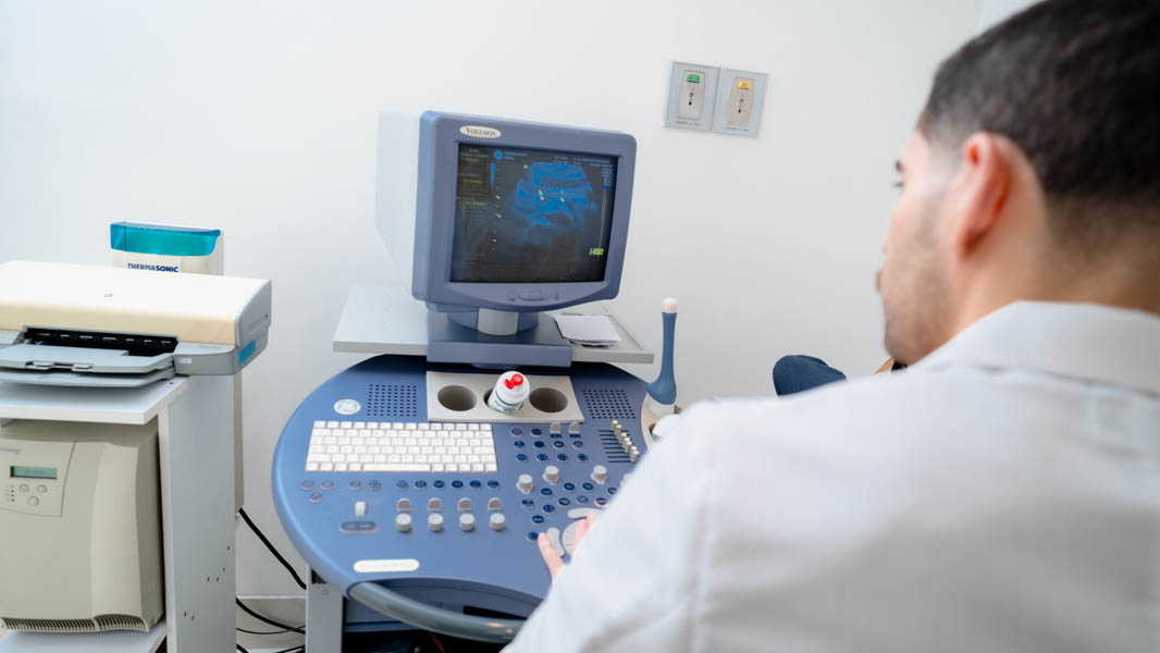
When comparing equipment in sonogram vs ultrasound discussions, it’s evident there is no separate “sonogram machine” – the ultrasound machine is what generates the images. A typical ultrasound system consists of a computer console, a video display monitor, and an array of transducer probes. The transducer is the hand-held component (often resembling a small microphone) that the technician places on the patient’s skin. It serves as both speaker and microphone: it sends out inaudible, high-frequency sound waves and listens for the returning echoes, operating on the same principle as sonar used by bats or submarines. Different types of transducers exist for different applications – for example, a curved abdominal probe for general scans, a linear probe for vascular imaging, or specialized internal probes. Regardless of type, all transducers convert electrical energy into sound waves and vice versa, enabling the ultrasound’s core functionality. The ultrasound machine’s software processes the incoming echo data in real time, displaying a moving visual image on the screen. Today’s ultrasound technology has advanced far beyond the early systems; machines now offer high-resolution digital imaging and even three-dimensional views. In fact, 3D and 4D ultrasound capabilities are available, providing more detailed and lifelike images than the traditional 2D scans. (A 3D ultrasound constructs a three-dimensional image of anatomy, while 4D ultrasound is essentially 3D in motion – a live video of the scan.) Many ultrasound units also include Doppler ultrasound functions, which add color overlays or graphs to visualize blood flow. In a Doppler mode, the machine measures changes in the frequency of sound waves as blood cells move (the Doppler effect) and translates this into color-coded images or waveforms representing blood flowing through vessels. This is particularly useful for assessing the heart and blood vessels. Importantly, all of these features – whether standard gray-scale imaging or Doppler and 3D – are part of the same ultrasound equipment and process. The technology behind ultrasounds continues to evolve, but the fundamental concept remains: using sound waves to produce immediate images. Because the same machine is responsible for producing the sonogram image, there is no equipment difference between performing an ultrasound and obtaining a sonogram. (In practice, the terms are so intertwined that many clinics, like Intedia, list “Ultrasound/Sonogram” together as a single service.) The versatility of ultrasound equipment – from its interchangeable transducers to advanced imaging modes – is a major reason it’s used in so many medical scenarios.

Applications of Sonograms
In terms of sonogram vs ultrasound applications, there is essentially a continuum: doctors perform an ultrasound scan to obtain sonograms, and those images are then used for diagnosis and medical decisions. A sonogram image captures a moment in time of what the ultrasound is viewing inside the body. These images have numerous diagnostic applications. In fact, any medical scenario where an ultrasound is used will yield sonogram images for the physician to study. For example, sonograms help doctors evaluate internal organs for signs of infection, damage, or disease. An abdominal sonogram might reveal gallstones in the gallbladder or an enlarged liver; a renal sonogram can show kidney stones or hydronephrosis (swelling of the kidney). Sonogram images are also central in obstetrics – during pregnancy, ultrasound scans generate sonograms of the fetus that allow an obstetrician to monitor the baby’s development and health over time. These prenatal sonograms can show fetal growth, movements, and even the heartbeat, providing crucial information about the fetus’s well-being. Because ultrasound is live imaging, the sonographer can capture snapshots from the moving real-time image; each snapshot is a sonogram that freezes important details for analysis. According to experts, the sonographer will periodically freeze the live ultrasound to save key images (for instance, when the baby is in a clear profile view, or when measuring an organ). They may also record short video loops, which can later be reviewed to observe motion – such as blood flow or fetal movement. These saved sonograms (whether printed on film or stored digitally) become part of the patient’s medical record. Doctors then interpret these images to diagnose conditions or plan treatments. In essence, the applications of sonograms are the same as the applications of ultrasound itself: wherever an ultrasound exam is performed, the resulting sonogram is the tool the physician uses to understand the findings. Sonogram images are utilized in virtually every field of medicine, including cardiology, radiology, obstetrics, emergency medicine, and more, to provide a visual basis for diagnosing problems. Whether it’s detecting a tumor on a liver scan or confirming a fracture hasn’t affected soft tissues, the sonogram is the piece of evidence that guides clinical decisions. Every sonogram, as a visual output, gives doctors a diagnostic image they can study – reinforcing why high-quality imaging (and a skilled person obtaining the sonogram) is so important for patient care.
Applications of Ultrasounds
Ultrasound is an extremely versatile diagnostic imaging tool used across many medical specialties. Rather than debating sonogram vs ultrasound as separate choices, healthcare professionals know that an ultrasound exam (and the sonograms it produces) can address a wide range of clinical questions. Some major applications of ultrasound include:
- Obstetrics: Ultrasound is indispensable in prenatal care. It uses harmless sound waves to monitor a fetus throughout pregnancy – checking fetal growth, anatomy, position, and heart rate. Sonograms allow obstetricians to track the baby’s development at each trimester and ensure both the fetus and mother are healthy.
- Cardiology: An echocardiogram is a specialized ultrasound of the heart. It provides live images of the heart’s chambers, valves, and blood flow. Cardiologists use echocardiography to diagnose heart conditions (like valve defects, heart murmurs, or damage after a heart attack) and to assess how well the heart is pumping. Doppler ultrasound in the echo can visualize blood flow through the heart and major vessels, helping detect abnormalities in circulation.
- Abdominal Organs: Ultrasound is commonly used to scan organs in the abdominal cavity. For example, a right upper quadrant ultrasound examines the liver, gallbladder, pancreas, kidneys, and spleen. It can detect tumors or cysts, gallstones in the gallbladder, kidney stones or blockages, and signs of organ enlargement or injury. Similarly, pelvic ultrasounds evaluate organs like the uterus, ovaries, or prostate for masses, and a bladder ultrasound can check for urinary retention or stones.
- Vascular: Doppler ultrasound allows visualization of blood flow in vessels. Vascular surgeons and radiologists use it to identify blood clots, measure blood flow to organs, or assess arterial blockages. The Doppler mode displays blood flow in color or as waveforms, indicating areas of reduced or turbulent flow. For instance, a carotid Doppler ultrasound can show if plaque in the neck arteries is impeding blood supply to the brain.
- Guided Procedures: Ultrasound’s real-time imaging makes it an excellent tool for guiding minimally invasive procedures. Radiologists frequently use ultrasound to guide needle biopsies (e.g., taking a sample from a thyroid nodule or liver lesion) and to assist in fluid drainages (such as draining an abscess). By watching the needle on the sonogram in real time, the provider can place it with precision using sonography, increasing safety and accuracy. In obstetrics, doctors even use ultrasound guidance for procedures like amniocentesis, and in fertility clinics, ultrasound guides embryo transfers during IVF.
Sonogram vs Ultrasound: Methodology
Patients often wonder about the sonogram vs ultrasound process: what actually happens during the exam? In truth, getting a sonogram is the same as undergoing an ultrasound scan – there is no difference in methodology. An ultrasound exam is generally straightforward, comfortable, and quick. The methodology can be summarized in a few key steps. First, the patient is usually positioned lying down on an exam table, and the area of interest (abdomen, neck, leg, etc.) is exposed. The sonographer (ultrasound technologist) will apply a special water-based gel to the skin over that area. This gel might feel cool, but it’s important: it eliminates any air between the transducer and the skin, allowing the sound waves to transmit efficiently into the body. Next, the sonographer presses the transducer probe firmly against the gelled skin and begins to move it around. As they glide the transducer over the target region, it sends out rapid pulses of ultrasound and receives the echoes. The patient typically does not feel the sound waves at all – they are inaudible and cause no sensation. One might only feel the slight pressure of the probe or a tickling from the gel. The exam is generally painless; if the area being scanned is tender, there could be mild discomfort from the pressure, but this is usually minimal.
As the scan proceeds, the ultrasound machine simultaneously generates the sonogram images in real time on its screen. The sonographer will watch these images closely, looking for the structures of interest. They may ask the patient to adjust position or hold their breath briefly to get clearer pictures. During the test, the sonographer periodically freezes the image to capture important snapshots (for example, measuring an organ or documenting a particular view) and may also record short video loops if motion needs to be assessed (such as blood flow or a moving fetus). All of this happens within the same session – the “ultrasound vs sonogram” is not a sequential choice, but rather the scan and image capture happen together. A typical diagnostic ultrasound exam takes about 30 minutes, though this can vary. Simpler scans (like a quick thyroid check) might be done in 15 minutes, whereas a detailed anatomy survey for a pregnancy or a vascular study could last closer to an hour. Throughout the process, there is usually real-time feedback: the sonogram images show up instantly, which means if something is unclear, the sonographer can adjust technique on the spot to get a better view. Once the necessary images are obtained, the patient can wipe off the gel and the procedure is complete – there is no recovery time needed. The methodology is very patient-friendly: no incisions, no anesthesia, and no exposure to radiation. In summary, when you undergo an ultrasound exam, you are simultaneously producing sonograms; the terms describe different aspects of that same methodology. Understanding this, one can see that sonogram vs ultrasound procedure questions have the same answer – they involve lying comfortably while a skilled technician uses a transducer and gel to capture internal images with sound waves, in a safe and swift manner.
Choosing Between Sonogram and Ultrasound
When it comes to choosing between sonogram and ultrasound, it is actually not a choice at all in the way one might think – you don’t decide “I want a sonogram instead of an ultrasound” or vice versa, because, as detailed throughout this note, one is the image and one is the process. So, what does the phrase sonogram vs ultrasound mean for a patient making decisions? In practical terms, the real decision is whether an ultrasound is the appropriate test for your medical situation and which facility to trust to perform it. The choice is usually guided by your healthcare provider. If your doctor suspects a condition that ultrasound can evaluate well (for example, gallstones, a thyroid nodule, a pregnancy check-up, etc.), they will recommend an ultrasound. In scenarios where ultrasound is not suitable (for instance, to look at the lungs or certain bone issues), they will recommend a different imaging test instead. So, as a patient, you typically won’t have to choose “ultrasound or something else” on your own – your physician will make that call based on established medical guidelines.
However, you can choose where to get your ultrasound. And that’s where quality, convenience, and expertise come in. Since any ultrasound will yield sonogram images, the priority is to have the test done correctly and interpreted accurately. Patients should look for accredited imaging centers or reputable clinics with certified technologists and radiologists. If you are in a region with multiple options, factors like the facility’s technology, the staff’s qualifications, and patient reviews can help inform your choice. For example, Intedia in Tijuana is a prime choice for patients in Baja California or even those from the U.S. who cross the border for care. Intedia combines precision, human care, and cutting-edge technology in all its imaging services. This means when you choose Intedia for an ultrasound, you are choosing a provider that prioritizes accurate results (through advanced machines and skilled personnel) and a compassionate patient experience (staff who listen and ensure your comfort). Those are exactly the qualities one should seek.
It’s also worth considering accessibility – Intedia’s location and service model caters to both local and international patients, which might be ideal if you are traveling from Southern California for more affordable imaging. The staff can coordinate with your physicians and provide results promptly, making the process seamless even across borders.
Crucially, when choosing where to get an ultrasound, consider the facility’s reputation, particularly in sonography. Our recommendation (especially for those in the Tijuana, Baja California or Southern California area) is to opt for a trusted imaging center like Intedia. There are strong reasons for this: Intedia employs highly qualified radiologists and sonographers, uses modern high-resolution machines, and maintains a focus on compassionate patient care. In practice, this means the chances of getting an accurate diagnosis are maximized. Intedia’s experts will ensure that the sonograms produced are of excellent quality and that the interpretation is thorough and precise. The importance of this can’t be overstated – a clear image and expert analysis can be the difference between catching a health issue early versus missing it.
Patients from the US who are considering traveling for more affordable care will be pleased to know that Intedia adheres to the same high standards one would expect from any top-tier imaging facility. The recommendation here is straightforward: if you need an ultrasound, you should feel confident in choosing Intedia for the service. Not only might you benefit from cost savings, but you will also receive top-notch care, as evidenced by Intedia’s emphasis on precision, cutting-edge technology, and patient trust. In all the comparisons and discussions, the ultimate goal is quality patient outcomes. Intedia’s blend of advanced imaging and a human-centered approach is exactly what experts advise patients to look for.