


What Is an Ankle MRI and Why Do Doctors Recommend It?
An ankle mri is one of the most useful tools doctors have when they need to get a clear picture of what is happening inside your ankle. Unlike an X-ray, which mainly shows bones, this type of imaging uses magnetic fields and radio waves to create detailed images of all the structures inside the ankle, including tendons, ligaments, cartilage, and bone tissue. If you have had ankle pain that does not go away, swelling that keeps coming back, or an injury that seems more serious than a simple sprain, your doctor may order this type of scan to find out exactly what is going on.
The images produced by this scan are so detailed that doctors can spot problems that would be completely invisible on other types of imaging. MRI is the preferred method for evaluating soft tissue injuries in the ankle because it captures structures like tendons and ligaments with exceptional clarity. This makes it the gold standard for diagnosing a wide range of ankle conditions.
How the Scan Actually Works
During the procedure, you lie down on a table that slides into a large machine shaped like a tube. The machine creates a strong magnetic field around the ankle area, and a computer uses the information gathered to build detailed cross-sectional images. The process is completely painless and does not involve any radiation, which is one of the main reasons doctors prefer it for certain types of injuries. A standard scan usually takes between 30 and 60 minutes, depending on the number of images needed.
Who Should Consider Getting This Type of Scan?
Not every ankle problem requires advanced imaging, but there are specific situations where a detailed scan is the only way to get a definitive answer. If you have experienced any of the following, your doctor may strongly recommend this type of evaluation:
- Persistent ankle pain that has not improved after weeks of rest and basic treatment
- A known or suspected ligament tear that may require surgery
- Tendon problems, including partial or complete ruptures
- Unexplained swelling or instability in the ankle joint
- Bone-related issues such as stress fractures that X-rays cannot clearly detect
- Cartilage damage or osteochondral lesions inside the joint
Common Ankle Injuries Diagnosed With MRI Imaging
The ankle is one of the most frequently injured joints in the human body. It supports your entire body weight with every step you take, and it is exposed to significant stress during physical activity. An ankle mri can identify a broad range of injuries with high accuracy, helping doctors make better treatment decisions and helping patients avoid unnecessary surgeries or prolonged incorrect treatments.
Ligament Tears and Sprains
The most common ankle injury is a sprain, which happens when one or more ligaments are stretched or torn. The anterior talofibular ligament, located on the outer side of the ankle, is the most frequently damaged ligament in the body. While many sprains heal on their own with rest and physical therapy, more severe cases involving complete ligament tears require a precise diagnosis before treatment can begin. MRI imaging can show exactly which ligament is affected, how severely it is damaged, and whether there are any other associated injuries in the surrounding tissue.
According to the American Academy of Orthopaedic Surgeons, chronic ankle instability affects a significant portion of people who experience lateral ankle sprains, which is why accurate imaging is essential for planning the right treatment approach.
Tendon Injuries in the Ankle
The ankle is surrounded by several important tendons that work together to allow movement and provide stability. The Achilles tendon, the largest tendon in the body, is particularly vulnerable to injury. Posterior tibial tendon dysfunction, peroneal tendon tears, and flexor hallucis longus tendon injuries are also common problems that can cause significant pain and functional limitations. An ankle mri provides a clear view of each of these tendons, allowing doctors to identify inflammation, partial tears, complete ruptures, and degenerative changes that might be causing your symptoms.
Bone Fractures That X-Rays Miss
Stress fractures are tiny cracks in the bone that develop gradually over time due to repetitive impact. They are especially common in athletes and people who suddenly increase their activity level. The problem with stress fractures is that they often do not show up on standard X-rays, especially in the early stages. MRI imaging is highly sensitive to these types of bone injuries and can detect them weeks before they become visible on conventional imaging. This early detection is crucial because continuing to put weight on a stress fracture can cause it to worsen significantly.
Understanding the Different Structures Visible on an Ankle MRI
One of the greatest advantages of this type of imaging is the sheer amount of information it provides in a single scan. A single ankle mri can show doctors the condition of bones, cartilage, tendons, ligaments, joint fluid, and surrounding soft tissue all at once. Understanding what the scan is looking at helps patients better appreciate why this test is so valuable.
Bone and Cartilage Evaluation
The ankle joint involves three main bones: the tibia, the fibula, and the talus. MRI imaging can detect subtle bone marrow changes, small fractures, cysts, and signs of infection or tumors that other imaging methods would miss entirely. Cartilage, which covers the surfaces of these bones and allows them to move smoothly against each other, is also clearly visible. Osteochondral defects, which are areas where the cartilage and underlying bone have been damaged, are a frequent source of chronic ankle pain and are best diagnosed through this type of scan.
Ligament Assessment in Detail
The ankle has multiple ligament complexes, including the lateral ligament complex on the outside of the ankle and the deltoid ligament on the inside. Each of these ligaments can be individually evaluated for thickness, continuity, and signal changes that indicate injury or degeneration. Knowing the specific condition of each ligament is critical when a surgeon is planning a repair or reconstruction procedure.
Posterior Ankle Structures
The posterior aspect of the ankle contains some of the most important and frequently injured structures, including the Achilles tendon and the posterior tibial tendon. Posterior ankle impingement syndrome, which causes pain at the back of the ankle during activities that require pointing the foot downward, is another condition that is best evaluated through detailed imaging. Accurate diagnosis of posterior ankle conditions significantly improves treatment outcomes and reduces the likelihood of chronic pain.
How to Prepare for Your Ankle MRI Appointment
Preparing for this type of scan is relatively straightforward, but there are some important things to keep in mind before your appointment. The magnetic field used during the procedure is extremely powerful, which means that any metal objects in or on your body need to be accounted for before you enter the scanning room.
What to Tell Your Doctor Beforehand
Before scheduling your scan, make sure your doctor knows about any of the following:
- Metal implants such as joint replacements, screws, or plates in any part of your body
- A pacemaker or any other electronic medical device
- History of working with metal, such as welding, where small metal particles could be present in your body
- Pregnancy or suspected pregnancy
- Claustrophobia or anxiety about enclosed spaces
- Any allergies, especially if contrast dye is going to be used
What to Expect on the Day of the Scan
On the day of your appointment, you will be asked to remove any jewelry, watches, or metal accessories. You may be given a gown to change into, or you may be able to keep your own clothing if it does not contain any metal. A technician will guide you through the entire process and answer any questions you have. If you are nervous about being in the machine, it is perfectly fine to ask your doctor about mild sedation options beforehand. The ankle is usually placed in a specialized coil that helps capture higher quality images of that specific area.
With or Without Contrast Dye?
Some ankle scans are performed with an injected contrast agent, which is a type of dye that makes certain structures stand out more clearly on the images. This is particularly useful when doctors are looking for infections, tumors, or inflammatory conditions. In other cases, the scan is performed without contrast because the standard images provide enough detail for diagnosis. Your doctor will decide which approach is best based on your specific symptoms and suspected diagnosis.
Reading Ankle MRI Images: What the Results Mean
After your scan is complete, a radiologist will examine the images and write a detailed report describing everything they observe. This report is then sent to your treating doctor, who will explain the findings to you and discuss the next steps. Understanding some of the common terms used in these reports can help you have a more informed conversation with your healthcare provider.
Common Findings and What They Indicate
Radiology reports for ankle scans often include terms that can sound alarming but actually describe conditions that are very treatable. Here are some common findings and what they typically mean:
- Tendinopathy: This refers to degeneration or chronic irritation of a tendon, often caused by overuse. It is not the same as a tear and is usually treated with physical therapy and rest.
- Partial tear: This means that some but not all of the fibers in a tendon or ligament are damaged. Treatment depends on the severity and location of the tear.
- Complete rupture: This means the structure has been entirely torn through and may require surgical repair.
- Bone marrow edema: This is swelling within the bone itself and is often a sign of a stress fracture, impact injury, or early bone disease.
- Joint effusion: This is excess fluid inside the joint, which is a sign of inflammation or injury.
- Osteochondral lesion: This refers to damage to both the cartilage and the underlying bone on a joint surface.
When a Second Opinion Is Worthwhile
Because MRI interpretation requires significant expertise, and because subtle findings can sometimes be missed or misinterpreted, getting a second opinion on your images can be valuable in complex cases. This is particularly true if your symptoms do not match the initial report findings, or if surgery is being recommended based on the results.
MRI Versus Other Imaging Options for Ankle Injuries
When you visit a doctor for ankle pain or an ankle injury, they have several imaging tools available to them. Understanding why an ankle mri is often chosen over other options helps clarify the value of this type of scan.
X-Rays and Their Limitations
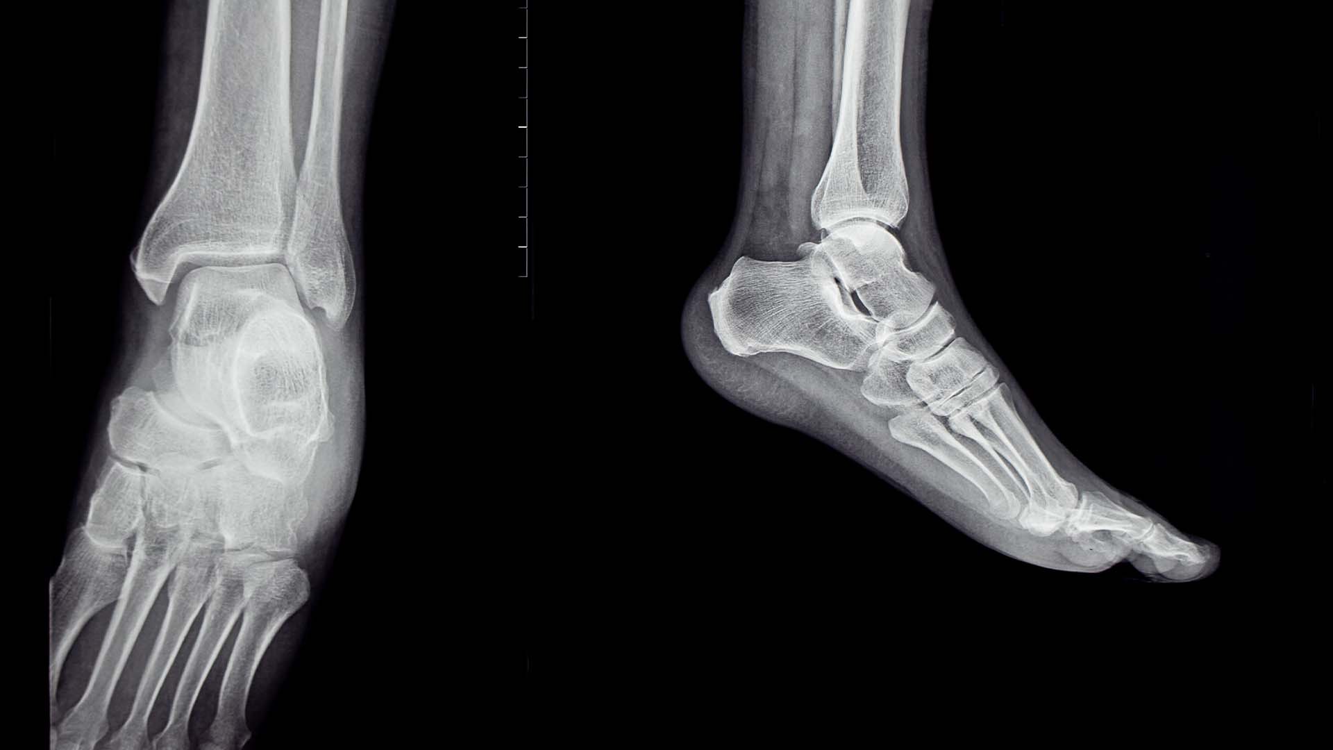
X-rays are almost always the first imaging test performed for ankle injuries because they are fast, inexpensive, and widely available. They are excellent for identifying obvious fractures and dislocations. However, X-rays cannot show soft tissue structures at all, which means they completely miss injuries to ligaments, tendons, and cartilage. They also struggle to detect subtle bone injuries like stress fractures in the early stages. For any suspected soft tissue problem, X-rays simply do not provide enough information.
Ultrasound as a Complementary Tool
Ultrasound is a real-time imaging technique that uses sound waves to create images of soft tissue structures. It is particularly useful for evaluating tendons and can even be performed while the ankle is moving, which helps identify dynamic instability. However, ultrasound is highly dependent on the skill of the operator and has limitations when it comes to viewing deep structures or the interior of joints. It is often used alongside MRI rather than as a replacement for it. According to the British Journal of Sports Medicine, combining ultrasound with MRI assessment provides the most comprehensive evaluation of complex ankle tendon injuries.
CT Scans and Bone Detail
Computed tomography, or CT scanning, provides detailed three-dimensional images of bone structures. It is particularly useful for complex fractures where the exact position of bone fragments needs to be understood before surgery. However, CT scans expose patients to radiation and do not show soft tissue structures as clearly as MRI. In most cases where a full evaluation of the ankle is needed, MRI remains the preferred choice.
Ankle MRI for Athletes: Performance and Recovery Implications
Athletes are among the most frequent users of ankle imaging because they experience a disproportionately high rate of ankle injuries. The demands placed on the ankle during sports like basketball, soccer, running, and gymnastics are enormous, and the consequences of an undiagnosed or mismanaged injury can be career-altering.
Diagnosing Sports-Specific Ankle Injuries
Certain injuries are particularly common in athletes and require very specific diagnoses to manage effectively. Syndesmotic injuries, which involve the ligaments connecting the tibia and fibula above the ankle joint, are a good example. These injuries are often called high ankle sprains and can take much longer to heal than standard lateral ankle sprains. Without proper imaging, they are frequently underdiagnosed. An ankle mri allows doctors to assess the severity of syndesmotic disruption and determine whether surgical fixation is needed.
Peroneal tendon subluxation, where the tendons on the outer side of the ankle slip out of their normal groove, is another condition that is best diagnosed through MRI. Athletes in sports that involve repetitive cutting and pivoting movements are particularly prone to this type of injury.
Planning Return to Sport After Injury
One of the most practical uses of ankle imaging in sports medicine is helping determine when an athlete is truly ready to return to full activity. Imaging can show whether a damaged structure has healed sufficiently to withstand the demands of training and competition. This objective information is invaluable for athletes, coaches, and medical teams trying to make safe and evidence-based return-to-play decisions.
Monitoring Chronic Conditions Over Time
For athletes with chronic ankle conditions, repeat imaging over time can show whether a condition is progressing, improving, or remaining stable. This helps doctors adjust treatment plans as needed and gives athletes a clearer picture of their long-term joint health. Longitudinal imaging studies have significantly improved understanding of how ankle injuries evolve and respond to different treatment approaches.
Finding the Right Place to Get Your Ankle MRI Done
Not all imaging centers are created equal. The quality of the equipment, the expertise of the technicians performing the scan, and the experience of the radiologists interpreting the images can all have a significant impact on the usefulness of the results you receive. When choosing where to have your scan performed, it is worth taking the time to research your options carefully.
What to Look for in an Imaging Center
Several factors distinguish high-quality imaging centers from mediocre ones. Consider the following when making your decision:
- The strength of the MRI machine, measured in Tesla. Higher field strength machines, such as 3T systems, produce sharper and more detailed images than older 1.5T machines, which can be particularly important for evaluating small structures in the ankle.
- The availability of dedicated musculoskeletal radiologists who specialize in reading joint and soft tissue images.
- The use of dedicated ankle coils, which are specialized attachments that significantly improve image quality for ankle scans specifically.
- Short wait times and flexible scheduling, since prolonged delays in diagnosis can affect recovery outcomes.
- Clear communication of results directly to patients and their referring physicians.
Why Intedia Stands Out for Ankle Imaging
If you are looking for a reliable and professional option for your imaging needs, Intedia offers high-quality diagnostic imaging services with a focus on accuracy and patient comfort. Their approach combines advanced technology with experienced professionals who understand the complexity of musculoskeletal conditions. For patients dealing with ankle injuries or persistent ankle pain, having access to a trustworthy imaging provider makes a real difference in how quickly and accurately they can get answers.
The team at Intedia is equipped to handle a wide range of imaging requests, including detailed ankle scans that require the kind of precision that makes a difference in diagnosis and treatment planning. Their commitment to quality imaging means you can trust the results you receive.
Treatment Options That Follow an Ankle MRI Diagnosis
Once you have a clear diagnosis from your imaging results, the path forward becomes much more defined. The treatment options for ankle conditions vary widely depending on the specific injury, its severity, and the individual patient’s health and activity level.
Non-Surgical Treatment Approaches
The majority of ankle injuries, even those that look significant on imaging, can be treated without surgery. Common non-surgical approaches include:
- Physical therapy focused on strengthening the muscles around the ankle to compensate for ligament or tendon damage
- Immobilization through bracing or casting to allow acute injuries to heal
- Anti-inflammatory medications to reduce pain and swelling
- Corticosteroid injections for targeted inflammation control in specific structures
- Platelet-rich plasma therapy, which is increasingly used for tendon and ligament injuries
- Activity modification to reduce stress on the injured structures during healing
When Surgery Becomes Necessary
Some conditions identified on an ankle mri do require surgical intervention. Complete tendon ruptures, significant osteochondral defects, and chronic ligament instability that has not responded to conservative treatment are common indications for surgery. The detailed information provided by MRI imaging helps surgeons plan the procedure precisely, which often leads to better outcomes and faster recovery times. Knowing exactly which structures are damaged and to what extent allows for a more targeted surgical approach.
Rehabilitation and Long-Term Care
Regardless of whether treatment involves surgery or conservative management, rehabilitation is almost always a critical part of recovery. A well-designed rehabilitation program rebuilds strength, restores range of motion, improves balance, and reduces the risk of re-injury. Follow-up imaging may be recommended at certain points during recovery to confirm that healing is progressing as expected. For patients who work with Intedia, having access to prompt follow-up imaging when needed is a practical advantage that supports better overall care.
Special Considerations for Ankle MRI in Children and Older Adults
Ankle injuries occur across all age groups, but the nature of those injuries and the way they appear on imaging can differ significantly between children, young adults, and older patients. Understanding these differences helps ensure that the right diagnostic approach is used for each patient.
Ankle Imaging in Pediatric Patients
Children have growth plates, which are areas of developing cartilage at the ends of the long bones. These growth plates are actually weaker than the surrounding ligaments, which means that what looks like an ankle sprain in a child may actually be a growth plate fracture. Standard X-rays may not always show these injuries clearly, making MRI an important tool for accurate diagnosis in younger patients. Additionally, because MRI does not involve radiation, it is particularly well-suited for children who may need imaging.
Ankle Imaging in Older Adults
In older adults, degenerative changes in the ankle joint are common and can complicate the interpretation of imaging results. Age-related tendon degeneration, osteoarthritis, and reduced bone density all affect how the ankle appears on a scan. According to the National Institutes of Health, the prevalence of ankle osteoarthritis increases significantly with age, and MRI can provide important information about the extent of joint degeneration that helps guide management decisions. Understanding the baseline condition of an older patient’s ankle helps doctors distinguish between acute injuries and pre-existing chronic changes.
Tailoring the Imaging Protocol
Modern imaging centers can adjust the specific sequences and parameters used during an ankle mri to optimize the images for different patient populations. Pediatric protocols may use faster sequences to minimize the time a child needs to stay still, while protocols for older patients may focus more heavily on bone quality and cartilage assessment. This flexibility makes MRI a highly adaptable tool for evaluating ankle conditions across the entire lifespan. Centers like Intedia that invest in up-to-date technology and trained personnel are better positioned to deliver this kind of personalized imaging care.