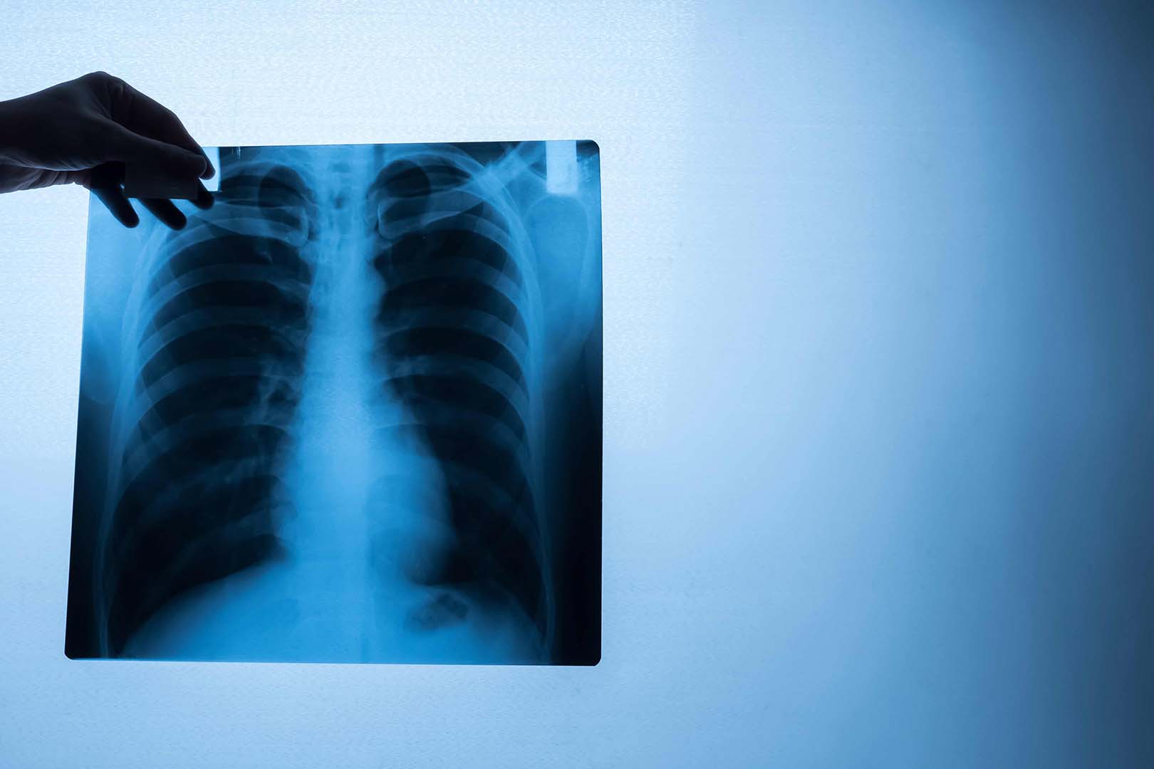
Preventing Pneumonia Complications Through Early Detection
Pneumonia is a serious lung infection that can become life-threatening if not addressed early. In fact, community-acquired pneumonia remains the leading infectious disease cause of death worldwide. Globally, pneumonia killed about 2.5 million people in 2023 alone. The infection inflames the air sacs in the lungs and can lead to dangerous complications like respiratory failure, sepsis, or fluid buildup around the lungs (pleural effusion). Early detection of pneumonia is critical because prompt treatment (often with antibiotics for bacterial cases) significantly improves outcomes. A pneumonia x ray – an imaging study of the chest – is one of the fastest and most reliable ways to detect pneumonia early and guide timely treatment. By catching pneumonia on an x-ray before it worsens, doctors can intervene sooner, helping prevent the infection from progressing to severe stages.
Early diagnosis not only saves lives but also reduces the risk of long-term complications. For example, pneumonia that is identified and treated early is far less likely to cause acute respiratory distress syndrome (ARDS) or lung abscesses. Medical experts emphasize that early detection and prompt antibiotic therapy are key factors in pneumonia survival. This is why healthcare providers often turn to chest x-rays at the first sign of pneumonia symptoms. Intedia, as a leading radiology and diagnostic center, is keenly focused on early detection – every pneumonia x ray at Intedia is performed with high-precision imaging technology and interpreted by specialized radiologists to ensure nothing is missed. By leveraging advanced digital x-ray systems, Intedia provides clear images of the lungs quickly, so that pneumonia can be confirmed or ruled out within minutes of the exam. This rapid clarity inspires confidence and enables physicians to start the right treatment immediately, helping patients avoid complications.
What is Pneumonia?
Pneumonia is an infection of the lungs that causes the air sacs (alveoli) to fill with fluid, pus, or cellular debris. In practical terms, pneumonia means part of your lung is inflamed and not functioning normally because of a germ – usually bacteria or viruses, and less commonly fungi – invading the lung tissue. As the infection spreads in the lung, it leads to consolidation, which is when the normally air-filled alveoli become solid with fluid/pus. Pneumonia can affect one lung or both lungs; an infection in both lungs is sometimes called “double pneumonia.” This illness ranges from mild to very severe, depending on the cause and the person’s overall health.
Common pneumonia signs and symptoms include cough (often producing phlegm or mucus), fever, chills, shortness of breath, and chest pain when breathing or coughing. Many patients also experience fatigue, sweating, or nausea. The classic description of bacterial pneumonia is a sudden high fever, chills, cough with thick discolored sputum, and sharp chest pain that worsens on inhalation (due to inflammation of the lung lining). Viral pneumonia symptoms may be more gradual and resemble a bad flu, while atypical pneumonia (like walking pneumonia caused by Mycoplasma) might cause milder symptoms despite an active infection.
Pneumonia is often categorized by where or how it was acquired. One major category is community-acquired pneumonia (CAP), which means the pneumonia developed outside of a hospital setting. CAP is the most common type and is frequently due to bacteria like Streptococcus pneumoniae (the pneumococcus). Another category is hospital or healthcare-acquired pneumonia, which occurs during a hospital stay and can involve more resistant bacteria. Regardless of the type, the mechanism is similar – germs enter the lungs (by inhalation of droplets or aspiration of mouth contents) and the immune system responds with inflammation, filling the alveoli with fluid. This fluid appears as white areas on a pneumonia x ray (more on that in the next sections).
Importantly, pneumonia is a lung infection that can affect anyone – from young children to the elderly. It tends to be more dangerous in infants, seniors, or people with weakened immune systems or chronic illnesses. If you suspect pneumonia (for example, if you have pneumonia signs like a persistent fever, productive cough, and chest pain), you should seek medical evaluation promptly. Doctors will typically perform a physical exam (listening for crackling sounds in the lungs) and likely order a chest x-ray to confirm the diagnosis.
How X Rays Help Diagnose Pneumonia
A chest x-ray is the primary imaging tool for diagnosing pneumonia, and it plays a central role in confirming the presence of a lung infection. On an x-ray image, healthy lungs appear mostly dark (because they are filled with air which X-rays pass through), whereas areas of pneumonia show up as white or gray opaque patches due to fluid/pus replacing the air. This contrast makes it relatively straightforward for a radiologist or physician to spot pneumonia on the film. In fact, the chest x-ray has long been considered the gold standard for initial pneumonia diagnosis. It allows doctors to see inflammation in the lungs, verify that pneumonia is present, and assess its extent and location.
When a patient has symptoms like cough, fever, and trouble breathing, a pneumonia x ray is often one of the first tests done. The x-ray can provide evidence of pneumonia within minutes, helping distinguish pneumonia from other issues such as bronchitis or asthma which might not show visible changes on imaging. Doctors rely on the x-ray to detect lung consolidation (solid-appearing lung tissue) that confirms pneumonia. According to medical experts, “the gold clinical standard for diagnosing pneumonia has been the chest X-ray to see if there is inflammation in the lungs.”. Secondary tests (like blood tests or pulse oximetry) can support the diagnosis, but the chest radiograph is key in visualizing the infection.
Beyond simply confirming the presence of infection, a pneumonia x ray helps in gauging severity and guiding treatment. The x-ray can show how much of the lung is affected – for example, a small localized pneumonia versus a widespread infection in both lungs. It can also reveal complications such as a pleural effusion (fluid around the lungs) or lung abscess. For instance, if an x-ray shows a large area of opacity covering an entire lobe, it indicates a significant lobar pneumonia that might require aggressive treatment and possibly hospital care. On the other hand, a modest patchy pneumonia in one segment might be managed with oral antibiotics at home. Thus, the chest x-ray findings help clinicians decide on hospitalization, antibiotic selection, and whether further tests are needed.
Another way x-rays aid diagnosis is by excluding other conditions. Sometimes symptoms of pneumonia can overlap with other illnesses like heart failure or lung cancer. An x-ray might reveal that the cause of symptoms is fluid overload in the lungs (pulmonary edema) rather than an infection, or it might uncover a tumor masquerading as pneumonia. While an X-ray alone cannot always give a definitive answer (for example, it cannot identify the specific bacteria causing pneumonia, and it cannot confirm cancer), it narrows down the possibilities and often clearly shows pneumonia patterns distinct from other problems. If the x-ray is ambiguous, doctors may follow up with a CT scan for more detail, but in the vast majority of cases, the plain chest x-ray is sufficient to diagnose pneumonia.
In summary, x-rays are indispensable in pneumonia diagnosis. They provide a fast, accessible, and highly informative look at the lungs. Clinics and hospitals around the world use chest x-rays as the first-line diagnostic imaging for suspected pneumonia because it reliably shows the infection (if present) and helps differentiate pneumonia from other causes of lung symptoms. By getting a pneumonia x ray early in your illness, you and your healthcare providers gain a clearer picture of what’s happening in your lungs, enabling prompt and appropriate treatment. Intedia’s radiology team recognizes the importance of this step – our general radiology service is optimized to deliver quick chest x-ray results with high clarity, so that pneumonia can be diagnosed without delay and managed before complications arise.
Key Features of a Pneumonia X Ray
What does pneumonia look like on an x-ray? The key feature is an area of increased density (whiteness) in the lung fields, reflecting the infected, fluid-filled lung tissue. Radiologists often use terms like “opacity,” “infiltrate,” or “consolidation” to describe these white patches on the x-ray. In a normal chest x-ray, the lungs appear mostly black because they are filled with air. When pneumonia is present, part of the lung will instead appear white or gray on the image, because air has been replaced by pus, fluid, and inflammatory cells. According to medical literature, “pneumonia appears as a white consolidation on a chest X-ray.” This white area may be large and uniform (as in lobar pneumonia) or scattered in smaller spots (as in bronchopneumonia), but either way it stands out against the darker background of healthy aerated lung.
A classic pneumonia x ray might show a homogeneous, cloud-like opacity in one lobe of the lung. For example, if the right middle lobe is infected, the x-ray will have a triangular white area roughly in the middle right lung zone. This consolidation can obscure normal borders between structures. Radiologists use the silhouette sign: when pneumonia in a lobe borders the heart or diaphragm, it can blur those normally sharp outlines. For instance, a right middle lobe consolidation often obscures the right heart border on the x-ray. Similarly, a pneumonia in the right lower lobe might make the edge of the diaphragm on that side indistinct. These clues help determine the location of the pneumonia on the image.
Another hallmark feature of pneumonia on x-ray is the presence of air bronchograms. Air bronchograms occur when the small airways (bronchi) remain air-filled and therefore show up as dark branching tubes running through the white opacity of consolidation. Seeing air bronchograms on an x-ray strongly suggests that the white area is indeed a lung consolidation (like pneumonia) rather than something like a pleural effusion which would obscure bronchi. In pneumonia, you might notice thin black lines within the white area – those are air-filled bronchi visible against the surrounding dense infection. This is a key finding radiologists look for.
The distribution of the opacity is also a key feature. Pneumonia can be localized to one segment or lobe (producing a focused opacity) or spread in a patchy way through multiple areas. A lobar pneumonia appears as a large contiguous white area occupying most of a lobe with clear edges at the fissures (the boundaries between lobes). In contrast, bronchopneumonia shows multiple patchy opacities of varying sizes in one or both lungs, often around the bronchi, giving a more mottled appearance. Interstitial pneumonia (often due to viral infections or atypical bacteria) might not show a classic consolidation at all, but instead a fine reticular (net-like) pattern or faint diffuse haziness throughout the lungs. We will discuss these patterns in more detail in the next section.
Understanding X Ray Terminology
Radiology reports for pneumonia can contain specialized terminology. Understanding these terms can help patients make sense of what a pneumonia x ray report means:
- Infiltrate: This is a general term indicating something has infiltrated (entered) the lung tissue. In the context of pneumonia, “infiltrate” usually refers to an area of opacity that may represent infection. For example, a report might say “right lower lobe infiltrate consistent with pneumonia.” It means they see a spot on the x-ray that likely corresponds to pneumonia in the right lower lung.
- Consolidation: As mentioned, consolidation means the lung’s air spaces are filled with liquid or solid material (pus, blood, fluid, cells), rendering that area solid on the x-ray. A radiologist might note “consolidation in the left upper lobe” – this is essentially confirming pneumonia in that region (assuming infection is the cause). Consolidation typically appears as a homogenous white area. If a consolidation is due to infection, it’s essentially the imaging word for pneumonia in that spot.
- Opacity or Density: These words describe any area on the x-ray that is more solid (whiter) than normal lung. “Opacity” is a catch-all term meaning an abnormal white area. Pneumonia opacities can be described by their shape or extent – for example, “patchy opacities in both lower lobes” suggests bronchopneumonia, whereas “wedge-shaped density in the right middle lobe” suggests lobar pneumonia. Opacity doesn’t specify cause; it could be pneumonia, fluid, tumor, etc. The context and associated signs (like air bronchograms) help determine that an opacity is likely a pneumonia consolidation.
- Air bronchogram: As explained above, this is when air-filled bronchi show up as dark lines within a lung opacity. If a report mentions “air bronchograms present,” it strongly supports the diagnosis of pneumonia or some kind of lung consolidation. Patients reading their report can take “air bronchogram” as a sign that the whitened area on the x-ray is indeed within the lung tissue (not outside it) and often points to pneumonia.
- Lobar vs. Patchy: Radiologists often describe distribution. “Lobar” means an entire lobe is involved – so you might read “lobar consolidation of the right upper lobe,” indicating a large pneumonia in that lobe. “Patchy” or “multifocal” means there are multiple spots in different areas – commonly used for bronchopneumonia or viral pneumonia (e.g., “patchy bilateral infiltrates”).
- Interstitial pattern: If the pneumonia is atypical or early, the x-ray might show an interstitial pattern – a diffuse, linear or grainy appearance instead of a big white blob. A report could say “interstitial infiltrates” which often correspond to pneumonia caused by viruses, Mycoplasma, or Pneumocystis pneumonia. Interstitial changes can look like a fine mesh across the lungs and can be subtle.
- Pleural effusion: This term might appear if there’s fluid accumulating around the lungs. Small effusions are not uncommon in pneumonia. The report might note “small right pleural effusion” meaning a bit of fluid has collected between the lung and chest wall on the right side, likely as a reaction to inflammation. Effusions appear as blunting of the costophrenic angle or a layering opacity on the side of the lung.
Understanding these terms can reassure you that the radiologist systematically checked the x-ray. For instance, a sample report could read: “Chest X-ray shows a dense consolidation in the left lower lobe with air bronchograms, consistent with pneumonia. Mild right pleural effusion noted. No discrete nodules or masses. Heart size normal.” In plainer language, this says there’s a pneumonia in the left lower lung, possibly some fluid on the right side, and no signs of tumor or heart failure. If anything in a report is unclear, patients should ask their doctor for clarification. But knowing that consolidation = pneumonia (in many cases) and air bronchograms = classic pneumonia sign can help decode the findings.
At Intedia, our radiologists aim to provide clear, jargon-free explanations alongside the official report. Every pneumonia x ray is interpreted by certified radiology specialists, and the results are communicated in a way that both doctors and patients can understand. Terminology is important for medical accuracy, but so is making sure the patient knows what their diagnosis means. We ensure that the key message – for example, that an x-ray shows pneumonia and not something else – is made clear. Our reports often include a conclusion like “Impression: Findings consistent with pneumonia in the right lung,” so you aren’t left guessing at complex terms. This commitment to clear communication, combined with top-notch imaging quality, is part of why Intedia stands out in diagnostic radiology services.

Common Patterns on Pneumonia X Rays
Not all pneumonias look the same on an x-ray. Doctors recognize several common patterns of pneumonia presentation in imaging, and each can hint at different causes or types of pneumonia. The main patterns often discussed are lobar pneumonia, bronchopneumonia, and interstitial pneumonia. Each of these terms refers to how the infection is distributed in the lungs and how it appears on the x-ray:
- Lobar pneumonia: This is a pneumonia that entirely (or almost entirely) involves one lobe of the lung. It’s characterized by a large, solid white consolidation in one lung lobe with relatively sharp boundaries at the fissures (the divisions between lobes). Lobar pneumonia is typically caused by “classic” bacteria such as Streptococcus pneumoniae. On a chest x-ray, lobar pneumonia produces a dense opacity that corresponds to an anatomical lobe – for instance, a right upper lobe pneumonia will show a consolidation in the top right portion of the lung field. This pattern often features air bronchograms and obscured normal landmarks (like the silhouette of the heart or diaphragm if the pneumonia is adjacent). We will delve more into lobar pneumonia specifics in the next section.
- Bronchopneumonia (Lobular pneumonia): This pattern is more patchy and widespread. Instead of one big lobe being white, you see multiple smaller areas of opacity scattered in one or both lungs. These opacities often originate around the bronchi and then spread to nearby alveoli – hence the name bronchopneumonia. On the x-ray, bronchopneumonia looks like patchy infiltrates – you might see cloudy spots in different segments, often more pronounced at the lung bases. The distribution can be bilateral and asymmetric, and it does not respect lobar boundaries as cleanly. Common causes include Staphylococcus aureus and atypical bacteria; it’s also seen following viral infections (secondary pneumonia). We will discuss bronchopneumonia in its own section as well.
- Interstitial pneumonia: This is usually caused by viruses or atypical organisms (like Mycoplasma pneumoniae, Chlamydia pneumoniae, or Pneumocystis in immunocompromised patients). Instead of classic consolidation, the infection primarily involves the lung interstitium – the tissue around the air sacs. On an x-ray, interstitial pneumonia manifests as a diffuse, reticular (network-like) or ground-glass pattern rather than a focal blob of opacity. It can look like faint streaks or a grainy texture across the lungs, sometimes described as “net-like infiltrates”. Often, these changes are subtle on x-ray and might be more apparent on a CT scan. Still, an experienced radiologist may see an interstitial pattern and suspect an atypical pneumonia. We will explore interstitial pneumonia in a dedicated section.
By recognizing these patterns, radiologists can sometimes infer the likely type of pneumonia. For instance, a lobar pattern in an adult with high fever and rusty sputum screams typical pneumococcal pneumonia. A diffuse interstitial pattern in a young adult with milder symptoms might suggest walking pneumonia due to Mycoplasma. That said, patterns can overlap and are not 100% specific – sometimes bronchopneumonia can coalesce into a lobar-looking consolidation, and severe viral pneumonia can have mixed interstitial and alveolar patterns. The pneumonia x ray is one piece of the puzzle, albeit a crucial one, in determining the pneumonia type.
In the sections below, we discuss Lobar Pneumonia, Bronchopneumonia, and Interstitial Pneumonia in detail, highlighting how each appears on x-ray and what it means for the patient.
Lobar Pneumonia
Lobar pneumonia is a classic form of pneumonia where an entire lobe of the lung is uniformly infected and filled with inflammatory exudate. On an x-ray, lobar pneumonia appears as a dense, homogeneous opacity occupying a large section of one lung, corresponding to a lobe’s anatomic boundaries. For example, if you have a right upper lobe pneumonia, the upper region of the right lung field will be white, often sharply demarcated by the horizontal fissure inferiorly. The rest of the lung outside that lobe remains clear (dark). This pattern gives lobar pneumonia a distinctive look – one lung region is “white-out” while adjacent areas are normal. The edges of the consolidation may form straight lines at the fissures. Lobar pneumonias usually also exhibit air bronchograms (air-filled bronchi within the dense lobe), reinforcing that it’s an alveolar filling process.
Clinically, lobar pneumonia often presents with more severe, acute symptoms: high fever, chills, productive cough with purulent (sometimes rust-colored) sputum, and pleuritic chest pain localized to the affected side. The classic organism is Streptococcus pneumoniae (pneumococcus), which historically causes lobar pneumonia affecting one lobe at a time. Other bacteria like Klebsiella can also cause lobar pneumonia (notably in upper lobes with potential cavitation in alcoholics), and some community-acquired pneumonias start lobar. In previously healthy individuals, lobar pneumonia is common.
On the x-ray, a lobar pneumonia can cause silhouette signs depending on the lobe:
- Right middle lobe pneumonia will obscure the right heart border (making that border disappear on the PA chest film).
- Right lower lobe pneumonia can blur the right diaphragm outline.
- Left upper lobe pneumonia might obscure the left heart border if it involves the lingula (the part of left upper lobe adjacent to the heart).
- Left lower lobe pneumonia can obscure the left diaphragm.
Radiologists use these clues to identify which lobe is involved even if the consolidation overlaps other structures. Additionally, lobar pneumonia does not usually cause volume loss – the lobe is filled with junk but not collapsed, so the lung size is maintained (or slightly enlarged due to inflammation). This differentiates it from lobar collapse (atelectasis), which would pull structures toward it. In lobar pneumonia, you might instead see bulging fissures in some cases (where the infected lobe is swollen and pushes fissures outward – classically described in Klebsiella pneumonia of the upper lobe).
Another feature sometimes seen in lobar pneumonia is the air-fluid level if an abscess forms, but that’s more of a complication than a defining feature. Generally, uncomplicated lobar pneumonias are solid opacities without cavities.
As the pneumonia resolves (with antibiotic treatment), x-rays will show the consolidation shrinking and breaking up over days to weeks, often clearing starting from the edges inward. Radiologists sometimes note “clearing” of a lobar pneumonia on follow-up films. It’s standard practice to get a follow-up chest x-ray about 6–8 weeks after a lobar pneumonia in older patients or smokers, to ensure it resolved completely and there isn’t an underlying mass that was hidden by the pneumonia. Studies have found a small percentage of lobar pneumonias can conceal tumors; approximately 2–9% of patients with pneumonia were later found to have lung cancer on follow-up imaging. So, clearing of the opacity is a good sign that it was purely an infection.
Bronchopneumonia
Bronchopneumonia represents a more scattered pneumonia infection that originates in the bronchi and spreads into the lung parenchyma in multiple areas. Instead of consolidating an entire lobe, bronchopneumonia causes patchy inflammation in lobules (small sections of the lung), often in multiple lobes simultaneously. On an x-ray, the hallmark of bronchopneumonia is multiple patchy opacities. These are typically small, ill-defined white patches distributed throughout one or both lungs, frequently bilateral. They tend to be more prominent at the bases but can be anywhere. The pattern might look like a sprinkling of cotton balls or clouds in the lungs rather than one big cloud.
A radiology report for bronchopneumonia might say “patchy bilateral infiltrates” or “multifocal pneumonia.” Unlike lobar pneumonia, the opacities of bronchopneumonia do not respect anatomical boundaries. They often cluster around airways – for example, you might see several patches around the bronchi in the lower lobes. Because the distribution is uneven, parts of the lung will appear normal while other parts have these patchy infiltrates. Air bronchograms may not be as obvious or may be absent in bronchopneumonia, especially if the patches are small – many of the infected areas are centered on bronchioles rather than entire alveolar regions, so the classic air bronchogram sign is less prominent (“No air bronchograms in many cases” of bronchopneumonia).
Common causes of bronchopneumonia include Staphylococcus aureus, Haemophilus influenzae, Pseudomonas (in hospital settings), and post-viral bacterial infections. It’s frequently seen in vulnerable populations like young children, the elderly, or people with chronic lung conditions, as well as a complication of influenza (flu) infections where Staph aureus causes secondary pneumonia in a patchy pattern.
Clinically, bronchopneumonia may present with a more gradual onset than lobar pneumonia. Symptoms can be variable – fever and cough are present, but sputum may be less or not localized, and the illness might be drawn out. Because the x-ray pattern is diffuse, it might be initially misinterpreted as something like atypical pneumonia or even pulmonary edema, depending on distribution. However, bronchopneumonia’s patchiness and asymmetry (not following a vascular pattern like edema does) give it away, as does the clinical context (patient has signs of infection).
On an x-ray, differentiating bronchopneumonia from, say, bronchitis is notable: acute bronchitis often does not show any consolidation on x-ray (it primarily affects airways, not alveoli), whereas bronchopneumonia will show these patchy infiltrates confirming it’s gone into the lung tissue. Thus, a pneumonia x ray that shows patchy infiltrates can validate that a person’s bronchial infection has progressed to pneumonia.
Treatment of bronchopneumonia involves antibiotics as well, but sometimes broader-spectrum coverage if a nasty organism is suspected (like Staph or gram-negatives, especially if hospital-acquired). The x-ray may lag behind in clearing – it’s not unusual for patchy opacities to take a few weeks to resolve on imaging, even if the patient feels better sooner.
From an imaging perspective, one feature of bronchopneumonia is that it can be multi-focal and recurrent. You might see improvement in some patches while new ones appear if the infection isn’t fully controlled, which is why follow-up imaging is done if patients don’t improve as expected. A follow-up pneumonia x ray after starting treatment should show those patches shrinking or gone; if not, doctors might reconsider the antibiotic choice or investigate other causes.
Interstitial Pneumonia
Interstitial pneumonia refers to pneumonia that primarily involves the lung interstitium – the network of tissue in the lung that includes the walls of the air sacs and the connective tissue supporting them – rather than filling the alveoli completely with pus. This pattern is often seen with atypical pneumonias (like those caused by viruses, Mycoplasma, Chlamydophila) or in certain pneumonias in immune-compromised patients (like Pneumocystis jirovecii pneumonia in HIV). On a chest x-ray, interstitial pneumonia does not show the classic lobar consolidation. Instead, it produces a diffuse, often subtle pattern of fine linear streaks or a grainy appearance, sometimes described as a “ground-glass” haze or reticular (net-like) pattern in the lungs.
In plain terms, an interstitial pneumonia x ray might look “busy” or blurry throughout the lungs, but without a big obvious white patch. The lung volumes are usually maintained, and you may see thickening of the lung markings everywhere. If mild, it can be easy to miss; the x-ray might even be read as “possible pneumonia, suggest clinical correlation” if findings are faint. Often, high-resolution CT is better at showing interstitial changes (revealing patterns like ground-glass opacities or small nodules), but the question is about x-rays, so we’ll focus on that.
One classic example is viral pneumonia (like influenza or RSV pneumonia): on x-ray it can show a diffuse bilateral infiltrate, sometimes more in the lower lobes, that is not dense or localized. It might be interpreted as interstitial edema vs pneumonia. Another example is Mycoplasma pneumoniae (walking pneumonia) – in many young patients, the chest x-ray shows a generalized haziness or fine patchy infiltrates; not the big lobar consolidation. Patients might not be extremely ill but have a lingering cough and fever.
Radiologically, some clues to interstitial pneumonia are:
- Reticular pattern: A kind of crisscross or net-like shadowing, indicating thickened interstitial lines.
- Peribronchial cuffing: This looks like fuzzy haloes around the bronchi, often from interstitial inflammation.
- Absence of air bronchograms: Because alveoli are not fully filled with fluid, we might not see distinct air bronchograms as in lobar pneumonia.
It’s important to note that early pneumonia of any cause can start with an interstitial appearance before consolidating. For instance, early pneumococcal pneumonia might show a faint interstitial pattern then evolve into a lobar opacity. So timing matters – a repeat x-ray a day or two later might “bloom” into a full consolidation if it’s typical bacteria. Conversely, if it stays interstitial, it suggests an atypical organism.
Pneumocystis pneumonia (PCP) in HIV/AIDS is a classic interstitial pneumonia – x-rays often show a bilateral interstitial infiltrate (ground-glass appearance) that can be subtle or progress to a “white out” if severe. Radiology texts note that PCP can present as a fine reticular pattern that was first attributed to something like edema, but persists and is actually infection.
Clinically, interstitial pneumonias often present with more gradual onset, dry cough (less sputum), lower fevers, etc. They’re sometimes called “walking pneumonia” because patients aren’t as acutely bedridden. However, viral pneumonias like COVID-19 or SARS can be interstitial and still cause severe illness – in those cases, chest x-ray shows diffuse haziness (though CT was heavily used for COVID to see ground-glass opacities).
On the report, a radiologist might phrase it as: “Diffuse bilateral interstitial infiltrates, consistent with atypical pneumonia. No focal consolidation identified.” That means they see evidence of pneumonia but not in the form of a lobar chunk. If you see “interstitial changes” in your x-ray report with suspicion of pneumonia, it implies an atypical or early infection.
Treatment for interstitial (atypical) pneumonia may differ (for example, macrolide antibiotics for Mycoplasma, antivirals for influenza, etc.), so identifying this pattern has implications for therapy. It’s one reason the term “atypical pneumonia” exists – because the x-ray and symptoms differ from typical lobar pneumonia, alerting clinicians to use different medications.
Comparing Pneumonia to Other Conditions
Chest x-rays can reveal a lot about lung health, but not every white patch on an x-ray is pneumonia. Other conditions can create lung opacities or symptoms that mimic pneumonia. It’s important for doctors (and reassuring for patients) to distinguish pneumonia from these other conditions to avoid misdiagnosis. Two conditions in particular often come up in the differential diagnosis of a suspected pneumonia on x-ray: lung cancer and tuberculosis (TB). We’ll discuss how each can be differentiated from pneumonia, both on imaging and clinically.
There are also other considerations – for example, how to tell pneumonia from heart failure (pulmonary edema) or from a non-infectious lung disease – but in this section, we focus on lung cancer and TB as they were specifically requested and are critical distinctions. In general, radiologists approach an opacity on x-ray with a mental checklist: Could this be pneumonia? Could it be a tumor? Could it be TB? The distribution, shape, and context help answer those questions.

Limitations of Pneumonia X Rays
While chest x-rays are incredibly useful, it’s important to understand their limitations in the context of diagnosing pneumonia. A pneumonia x ray provides a two-dimensional snapshot of the chest and has some constraints:
False Negatives (Pneumonia can be missed)
A normal chest x-ray does not always completely rule out pneumonia. Especially in the early stages of infection, an x-ray might appear clear even if the patient is symptomatic. There are a few scenarios where pneumonia might not show up on an initial x-ray:
- If the infection is very early or mild, the inflammatory changes might not yet be extensive enough to be visible.
- Dehydration can reduce the radiographic signs of pneumonia because the lack of fluid makes the consolidation less conspicuous. In a dehydrated patient, the usual fluffy infiltrate might be subtle or absent.
- Immunosuppressed or elderly patients might not mount a strong localized inflammatory response, so their pneumonias can be atypically subtle on x-ray. They might have pneumonia with very faint infiltrates or none at all initially.
- A very high respiration rate or inability to take a deep breath can make the x-ray less sensitive (the lungs not fully expanded on the film).
False Positives (Mimickers of pneumonia)
As discussed in the previous section, not everything that looks like pneumonia on x-ray is actually pneumonia. Scarring, tumors, fluid, or collapsed lung segments can all create opacities that might initially be interpreted as pneumonia. Radiologists use context and additional imaging to sort these out, but a non-expert viewing an x-ray might easily mistake, say, a tumor for a pneumonia. Even radiologists sometimes hedge until further tests confirm. For instance, atelectasis vs consolidation: a lobar collapse can look similar to a lobar pneumonia on x-ray. There are radiographic clues (volume loss, fissure shift for atelectasis vs none for pneumonia), but it can be tricky in a single image. So, an x-ray might “diagnose” pneumonia, but if the patient doesn’t respond to treatment, it could have been something else, meaning the x-ray alone wasn’t definitive.
Lack of Etiology Information
A chest x-ray cannot tell you what specific germ is causing the pneumonia. The appearance of pneumonia on x-ray is generally similar whether it’s caused by bacteria, viruses, or fungi. There are some patterns suggestive of certain pathogens (like bulging fissure in Klebsiella, or interstitial pattern for viral), but they are not reliable enough to guide exact treatment. For definitive identification, one needs lab tests (sputum culture, PCR, blood tests). The x-ray shows “there is pneumonia”, but not “this pneumonia is due to streptococcus” for example. So while x-ray guides the presence and extent of disease, doctors must use clinical judgement or additional tests to determine the cause and choose proper antibiotics or antivirals.
Given these limitations, what are the practical steps? If pneumonia is strongly suspected clinically but the x-ray is negative or equivocal, doctors may:
- Repeat the x-ray in 1-2 days (infiltrate might declare itself).
- Do a Chest CT scan which is much more sensitive to lung changes (a CT can see pneumonia that x-ray doesn’t, and can differentiate causes of opacities better).
- Use ultrasound at bedside for quick check if available (useful especially for peripheral pneumonias or differentiating pneumonia vs effusion).
- Treat presumptively and see if the patient improves (if they do, likely was pneumonia even if x-ray lagged).
At Intedia, we recognize these limitations and thus provide a comprehensive diagnostic approach. Our radiologists give detailed reports noting any uncertainties or alternative considerations. If there’s any doubt or something unusual, we work with referring physicians to arrange follow-up imaging (like a CT) promptly. By combining top-notch imaging with clinical insight, we minimize the impact of x-ray limitations. In essence, a pneumonia x ray is a powerful “snapshot” tool – but it’s one part of the bigger diagnostic picture.
Next Steps After a Pneumonia X Ray
So, you’ve had a pneumonia x ray and it confirms that you have pneumonia (or at least strongly suggests it). What comes next in your care? The chest x-ray finding is just the beginning of the management process. The next steps typically involve treatment, monitoring, and sometimes further testing to ensure the pneumonia is properly addressed and to check for any complications or underlying issues.
Initiation of Treatment
The primary next step is to start appropriate treatment for pneumonia. For most bacterial pneumonias, this means antibiotics. The choice of antibiotic will depend on the suspected type of pneumonia (community-acquired vs hospital-acquired, typical bacteria vs atypical, etc.) and patient factors (allergies, local resistance patterns). For example, a community-acquired pneumonia might be treated with a macrolide or doxycycline, or a respiratory fluoroquinolone, or a combination like beta-lactam + macrolide, based on guidelines. If the x-ray is very extensive (suggesting a severe pneumonia) or the patient has low oxygen levels, hospital admission might be necessary for IV antibiotics, oxygen therapy, and supportive care. Antibiotic therapy should ideally start as soon as possible after diagnosis – studies show prompt treatment improves outcomes.
Supportive Care and Symptom Management
Alongside specific medication, next steps include supportive measures:
- Rest and fluids: Patients are advised to rest and stay hydrated to help the body fight the infection and loosen secretions.
- Fever control and pain relief: Medications like acetaminophen or ibuprofen can reduce fever and alleviate chest pain from pleurisy. Chest pain (especially sharp pain with deep breaths) is common in pneumonia due to pleural inflammation; relieving this can help patients breathe deeper and cough more effectively.
- Cough support: If the cough is severe or causing discomfort, sometimes doctors recommend expectorants or inhalers. However, suppressing the cough too much isn’t ideal because you want to clear mucus. So often just staying hydrated and maybe using a humidifier is suggested, unless cough is so bad it’s causing vomiting or sleeplessness.
- Oxygen therapy: If the pneumonia has caused low oxygen levels (which might be indicated by shortness of breath or measured by a pulse oximeter/hospital test), supplemental oxygen will be given, either via nasal cannula or mask in the hospital. In very severe cases, mechanical ventilation might be needed (ICU care).
Further Testing (when needed)
In many straightforward cases, a pneumonia x ray and clinical exam are enough to start treatment. But sometimes additional tests are done:
- If the patient is hospitalized or if there’s concern for unusual organisms, they may send sputum samples for culture, do a blood culture, or specific antigen tests (like pneumococcal or Legionella urine antigen tests, flu swabs, etc.). These help identify the bug to tailor antibiotics.
- If there’s a significant pleural effusion seen on the x-ray, a doctor might order a ultrasound of the chest or even do a thoracentesis (insert a needle to draw fluid) to analyze it or relieve it. Infected fluid (empyema) might need chest tube drainage.
- For patients not responding to initial treatment or who have risk factors, a bronchoscopy might be considered to visually inspect the airways and get a sample from the lungs.
- As mentioned in limitations, if something about the x-ray is atypical (possible mass, etc.), a CT scan of the chest may be ordered next. CT can reveal details not visible on x-ray, like small abscesses, cavities, hidden tumors, or adenopathy. CT is also helpful if the patient isn’t improving, to look for complications like an abscess or empyema that might require a different approach.
Follow-Up Imaging
It’s common practice to do a follow-up chest x-ray some weeks after treatment to ensure the pneumonia has cleared. Follow-up is especially important for smokers and older patients because of the aforementioned possibility that an underlying tumor or other issue was hidden. Typically, 6-8 weeks after completing treatment, a repeat x-ray is done. If it’s clear, great. If residual opacity remains, further investigation is needed (like CT or bronchoscopy) to rule out something like cancer.
Doctors, however, generally do not do x-ray much sooner than that for follow-up, unless the patient is getting worse or not improving, because x-ray changes lag behind clinical improvement. You can feel better but still see some shadow on x-ray for a while. One study in Chest showed majority of pneumonia clears by 6 weeks, but a chunk needed 12 weeks, especially in older folks. So timing of follow-up can vary. If you’re younger and healthy, some doctors skip follow-up x-ray if you’re completely well, but many will still do it for completeness.
When things don’t go as expected
If after 48-72 hours of antibiotics the patient is not improving (fever not coming down, breathing still very difficult, etc.), that’s a trigger for re-evaluation. Next steps might include broadening antibiotic coverage (maybe the germ is resistant or atypical), repeating imaging to look for complications, or considering an alternate diagnosis (like pulmonary embolism or TB). For instance, if an initial outpatient x-ray showed pneumonia but the person worsens, a CT might reveal an abscess that needs drainage or a large effusion needing a chest tube.
Specialist Consultation
In complicated cases or if the patient has other issues, doctors might involve specialists. A pulmonologist (lung specialist) or an infectious disease specialist might be consulted if:
- The pneumonia is severe (ICU care).
- It’s not responding to standard therapy.
- There are unusual features (like possible TB or an immunocompromised host who could have Pneumocystis or fungal pneumonia).
- There’s a question of needing bronchoscopy or other interventions.
At Intedia, we facilitate the next steps by making sure the referring doctors get the imaging results promptly and any recommended follow-up is clearly indicated on the report. For example, our report might say: “Large right pleural effusion noted; ultrasound-guided thoracentesis could be considered” or “Recommend follow-up chest X-ray in 6 weeks to confirm resolution.” We can also connect referring physicians with our network of specialists if advanced imaging (like CT) shows something requiring special care. Essentially, once the pneumonia x ray is done, Intedia doesn’t just leave you with a film; we help navigate the subsequent steps through comprehensive reporting and advice.
In a typical scenario: The x-ray confirms pneumonia → Doctor starts antibiotics (e.g., a course of levofloxacin or amoxicillin-clavulanate + azithromycin for CAP) → You go home with instructions (rest, fluids, etc.) or stay in hospital if needed → You’re told to come back for follow-up in a couple days if no improvement, otherwise finish the medicine → You have a follow-up appointment in a week or two to ensure you’re better → In 6-8 weeks you get a repeat x-ray at Intedia to ensure it’s all clear. During that follow-up appointment, the doctor might also review any lab test results that came in (like culture results) and adjust treatment if necessary.
When to Consult a Specialist
Most cases of pneumonia, especially straightforward community-acquired pneumonia in an otherwise healthy person, can be managed by general practitioners or internists without issue. However, there are situations where involving a specialist – such as a pulmonologist (lung specialist) or an infectious disease (ID) specialist – is prudent. Knowing when to consult a specialist can impact recovery and help address complicated aspects of the disease.
You should consider seeing a specialist or ask your doctor about a referral if:
The pneumonia is severe or not improving as expected
If you have a severe pneumonia that landed you in the ICU or required advanced support (like a ventilator or chest tube for drainage), a pulmonologist will likely be involved in your hospital care. Pulmonologists have expertise in managing respiratory failure, complex imaging, and interventions like bronchoscopy. Similarly, an infectious disease specialist might be consulted in the hospital for severe pneumonia to ensure the antibiotic coverage is optimal, especially if the causative organism is unusual or resistant. If 48-72 hours have passed and you’re not responding to antibiotics, an ID specialist can help figure out if the bug might be atypical or if there’s another infection at play.
You have recurrent pneumonias or an underlying lung condition
If this is not your first pneumonia or if you have chronic lung diseases such as COPD, asthma, or bronchiectasis, a pulmonologist consultation is wise. Recurrent pneumonias in the same area may indicate an underlying problem that a pulmonologist could evaluate (like a tumor, bronchi blockage, or congenital issue). They can perform specialized tests like bronchoscopy to look inside the airways for any obstruction or take samples. For example, say you’ve had pneumonia twice in the right middle lobe in six months – a pulmonologist can investigate why that lobe is vulnerable (sometimes it could be anatomical like a narrow bronchus, or an aspirated foreign body, etc.).
There are complications that need special procedures
If your pneumonia led to a significant pleural effusion or empyema, a pulmonologist or sometimes a thoracic surgeon would be consulted to drain it. Pulmonologists can do thoracentesis and place chest tubes, and manage them. If a lung abscess formed, a specialist would manage it (sometimes via interventional radiology drainage or longer antibiotics guided by ID consult). Essentially, any complication beyond the scope of routine medicine calls for the appropriate specialist.
Suspicion of uncommon pathogens or need for tailored therapy
An infectious disease specialist is very helpful if the pneumonia is caused by an unusual organism (e.g., tuberculosis, fungal pneumonia like histoplasmosis or coccidioidomycosis, or in immunocompromised hosts Pneumocystis jirovecii pneumonia). They have expertise in these less common infections and the special medications required to treat them. For instance, treating TB is a long 6+ month process with multiple drugs – an ID doctor would coordinate that therapy. If a patient is not responding to standard antibiotics, an ID specialist might suggest advanced diagnostics (like antibody tests, bronchoscopy for cultures) to find a rarer cause.
Underlying immune issues
If someone gets pneumonia frequently or has one that’s very hard to cure, there might be an immune system issue. An ID specialist or immunologist might evaluate for immunodeficiencies (like checking Ig levels for something like CVID, or HIV testing if risk factors present). Sometimes a simple clue like recurrent unusual infections prompts a deeper immune work-up. Specialists can coordinate these tests and interpret them to see if prophylactic measures or long-term treatments are needed to prevent future infections.
Need for lung rehabilitation or follow-up
After a severe pneumonia, especially in older patients or those with pre-existing lung disease, lung function might be reduced. A pulmonologist can measure lung function via spirometry after recovery and see if there’s any persistent impairment that needs rehab or inhaler therapy. They can also ensure that any scarring hasn’t significantly impacted breathing capacity.
Preventive consultation
A specialist might also advise on preventive measures if you fall into certain categories. For example, after recovering from pneumonia, a pulmonologist or general doctor might refer you to get vaccinated (pneumococcal vaccines, annual flu shot, etc.). If you’re a smoker, they would strongly advise cessation and might refer you to smoking cessation programs. If aspiration was an issue, they might involve a speech-language pathologist to assess swallowing, or a gastroenterologist if reflux is causing aspiration.
From the patient perspective, how do you know if you need a specialist? Generally, trust your primary doctor’s guidance – they will refer when needed. But you should certainly ask about it or seek a second opinion if:
- After completing treatment, you’re still feeling unwell or have lingering respiratory issues.
- You’ve had multiple pneumonias and no one has investigated why.
- You have a complicated medical history (like HIV, or you’re on chemotherapy, etc.) which might predispose to unusual infections – an ID doctor’s involvement early can be beneficial.
- You feel your questions about the disease or its prevention aren’t fully answered – sometimes a specialist can provide more detailed counseling (e.g., an ID specialist discussing long-term prophylactic antibiotics or a pulmonologist discussing inhaler use to strengthen lungs post-pneumonia).
At Intedia, while we are primarily a diagnostic center, we have a robust network of specialists we work with. If our imaging raises suspicion of something requiring specialist care (say we see a possible mass on follow-up or a cavity that looks like TB), we will mention in our report that specialist evaluation is recommended. We can also fast-track referrals – for instance, if your pneumonia x ray suggests possible TB, we alert your physician and can help connect with a pulmonologist or ID specialist for further testing immediately.
Prevention and Early Detection Strategies
Recovering from pneumonia or hoping to avoid it in the first place naturally leads to the question: How can I prevent pneumonia, and catch it early if it does occur? There are several effective strategies, combining lifestyle measures, vaccinations, and timely medical evaluation, that significantly reduce the risk of pneumonia and its complications.
Vaccinations
This is arguably the single most powerful preventive tool. Several vaccines protect against organisms that commonly cause pneumonia:
- Pneumococcal vaccines: These target Streptococcus pneumoniae, the leading bacterial cause of pneumonia. There are two main types for adults, Pneumovax 23 and Prevnar 13, which cover different strains. Guidelines typically recommend adults over 65 receive both (in a scheduled sequence), as well as younger adults with risk factors (like chronic heart, lung, kidney disease, diabetes, or smokers). There are also pneumococcal vaccines as part of the routine childhood immunization schedule for kids (PCV13/PCV15 for infants, and PPSV23 for certain high-risk children).
- Influenza vaccine: The flu can directly cause viral pneumonia, and it also can weaken the lungs/immune system enough to get a secondary bacterial pneumonia. Annual flu shots have been shown to reduce the incidence of severe influenza and pneumonia. Flu vaccine is recommended for everyone over 6 months of age yearly.
- Other vaccines: Haemophilus influenzae type b (Hib) vaccine (for children) and Pertussis (whooping cough) vaccine (DTaP in kids, Tdap boosters in adults) also indirectly prevent pneumonia, as these organisms can cause respiratory infections and pneumonia. Also, Varicella (chickenpox) vaccine in children prevents chickenpox pneumonia (and by extension, the risk of varicella leading to bacterial pneumonia). Measles vaccine prevents measles, which can lead to severe pneumonia as a complication. Essentially, keeping up with the entire recommended immunization schedule throughout life protects your lungs.
In essence, vaccinations create a shield. While they don’t guarantee you’ll never get pneumonia, if you do contract an infection it’s likely to be milder. For example, a vaccinated person might get a less severe pneumonia or avoid hospitalization.
Good Hygiene and Infection Control
Pneumonia often starts from catching a cold or flu, then escalating. So, preventing respiratory infections in general helps:
- Hand washing: Regularly wash hands with soap and water, especially during cold/flu season or after being in public places. Our hands pick up germs and we often touch our face, transmitting them to nose/mouth.
- Avoid smoking and secondhand smoke: Smoking damages the respiratory tract’s defenses (like the cilia that sweep out germs), significantly increasing pneumonia risk. Quitting smoking is one of the best things you can do – it not only reduces pneumonia risk but improves overall lung function and immunity. Avoiding secondhand smoke is similarly important, especially for children.
- Limit alcohol intake: Heavy alcohol use can increase pneumonia risk by impairing the immune system and the reflexes that prevent aspiration (inhaling secretions into lungs). Moderation or avoidance is wise for many health reasons including pneumonia prevention.
- General infection avoidance: This includes avoiding close contact with individuals who are coughing or sick (if you’re vulnerable), and not sharing utensils or cups during cold/flu season. Covering your own coughs/sneezes and staying home when ill can protect others (this is a community strategy to reduce overall transmission).
- Dental hygiene: Believe it or not, poor oral hygiene can increase risk of aspiration pneumonia (especially in older folks) because harmful bacteria can colonize the mouth and then get aspirated. Regular dental care and oral hygiene can mitigate this.
Health Maintenance
Keeping your body’s defenses strong is a broad goal:
- Nutrition: A well-balanced diet supports immune function. Malnutrition (especially in older adults or those with chronic illness) weakens immunity, so ensure adequate intake of vitamins and proteins.
- Exercise: Regular physical activity can improve lung capacity and immune health. It doesn’t have to be intense – even walking most days can have benefits. Exercise also helps conditions like COPD patients clear secretions and improves overall stamina.
- Adequate sleep: Sleep deprivation can impair immune response. Aim for quality sleep which is when your body repairs and prepares immune cells.
- Managing chronic conditions: If you have diabetes, keep it controlled (high blood sugar can hamper immune cells). If you have asthma or COPD, follow your treatment plan (inhalers, etc.) to reduce baseline lung inflammation and risk of infection. For heart failure patients, managing fluid levels can avoid pulmonary edema which predisposes to pneumonia.
Early Detection and Prompt Treatment
Even with prevention, pneumonia can sometimes occur. Recognizing early signs and seeking prompt medical evaluation can make a difference in outcomes (the earlier pneumonia is treated, the quicker recovery and less chance for complications). Key pneumonia signs to watch for include:
- Persistent fever, often with chills.
- A cough that becomes productive (yellow/green or bloody phlegm).
- Chest pain when breathing or coughing (from pleurisy).
- Shortness of breath beyond what a normal cold would cause.
- Unusual fatigue or confusion (especially in older adults, confusion can be an early sign of infection).
If these occur, don’t delay in seeing a healthcare provider. A chest x-ray can confirm if it’s pneumonia. As we noted, sometimes pneumonia can present subtly, especially in elders (maybe just weakness and slight cough without a big fever). It’s better to check earlier than to wait until severe.
At Intedia, beyond providing diagnostic imaging, we support prevention and early detection in a few ways. Our radiology reports might include a note if we spot something like chronic changes (maybe evidence of smoking damage on the x-ray) reminding the physician and patient about smoking cessation. If a patient comes for frequent imaging due to recurrent respiratory issues, our team may gently suggest discussing vaccination or specialist consultation.
By following these prevention and early detection strategies, you greatly increase your chances of either avoiding pneumonia altogether or catching it at a very treatable stage. As the saying goes, an ounce of prevention is worth a pound of cure – keeping your lungs healthy and germs at bay is far easier than dealing with a full-blown pneumonia and its aftermath.
How to Prepare for a Pneumonia X Ray
If your doctor has ordered a chest x-ray because you have symptoms of pneumonia (or for a follow-up), you might wonder how to prepare and what to expect. The good news is that a pneumonia x ray is a quick, non-invasive, and generally straightforward procedure. Here are some tips and steps to ensure the process goes smoothly:
Clothing and Accessories
You will typically be asked to remove clothing from the waist up and change into a hospital gown. This is to eliminate any objects that might obstruct the x-ray image. Metal objects in particular can block the x-rays and show up on the image, so you’ll need to remove necklaces, bras with metal hooks, piercings, or any jewelry around the neck/chest area. It’s best to wear a simple shirt that’s easy to take off. If you have long hair, you might be asked to tie it up (especially if it has hairpins or metal clips) so that it doesn’t cover your back or chest.
Informing the Technician
Let the x-ray technologist know if you are or could be pregnant. This is crucial, as chest x-rays involve a small dose of radiation and although the dose is low (and usually directed away from the abdomen), special precautions or alternative imaging (like ultrasound) might be used for pregnant women to avoid any risk to the fetus. At Intedia, and most imaging centers, there are strict protocols about pregnancy – often they’ll have you sign a form confirming if you’re not pregnant. If you are pregnant and a chest x-ray is very necessary, they will use a lead apron to shield your abdomen.
Also, mention if you’ve had any recent barium contrast studies (like a barium swallow or upper GI series) or if you have any implants. Metallic implants (like pacemakers or rods) usually don’t interfere with chest x-rays much, but it’s good for them to know so they don’t misinterpret them as something pathological on the image.
During the X-ray
- You’ll stand (or sometimes sit) in front of the x-ray detector. For a pneumonia check, typically two views are taken: a posterior-anterior (PA) view, where you stand facing the detector (with the x-ray machine behind you shooting through your back to front), and a lateral view, where you stand sideways against the detector.
- The technologist will position you – often you’ll press your chest against the detector for the PA view, roll your shoulders forward or place your hands on your hips and elbows forward to move the shoulder blades off the lung fields. Don’t worry, they will guide you exactly how to pose.
- You’ll need to take a deep breath and hold it when the x-ray is taken. They usually say “Take in a deep breath, hold it” and then click, then “you can breathe.” Taking a deep breath inflates your lungs fully, giving a clearer picture and better contrast (more air makes the lungs appear darker, which makes any white pneumonia more conspicuous). Holding still is important because motion can blur the image. A chest x-ray exposure is very quick (fractions of a second), so the breath-hold is only for a moment.
- For the lateral view, you’ll turn sideways and often raise your arms above or behind your head to avoid your arm soft tissues overlapping the lung. Again, you’ll be asked to take a breath in and hold it briefly.
The whole x-ray exam takes maybe 5-10 minutes, mostly for positioning; the actual radiation exposure is milliseconds. It’s painless. You won’t feel anything from the x-rays themselves. The machine might make a brief noise when it takes the picture.
After the X-ray
There’s no special aftercare. You simply get dressed. There are no side effects – you won’t be radioactive or anything. At Intedia, images are digital, so they’re ready almost immediately for review. In an urgent situation, the radiologist can often read it within minutes. Otherwise, standard turnaround might be later that day. But for pneumonia, usually they expedite results if you’re waiting in clinic.
Comfort and Logistics
If you have difficulty standing (say you’re feeling very weak or lightheaded due to illness), inform the tech. Chest x-rays can be done sitting in a wheelchair or even lying down (though supine AP films are a bit less clear). Intedia’s radiology team is accustomed to working with patients who may not feel well, so they’ll help. If you’re coughing a lot, you might need a moment to compose yourself and they may need to clean the detector after if you cough on it – but no worries, that’s routine. They also often give you a lead shield to wear around your lower body for any stray radiation, though modern x-ray beams are very targeted.
Bring any previous imaging results if done elsewhere (for example, if you had an older chest x-ray or CT, sometimes comparisons are useful). If done at Intedia before, our radiologist will automatically compare with past images in our system.
Home vs Clinic
If you are extremely ill and cannot come to the imaging center, in some cases, pneumonia can be evaluated by a mobile x-ray service or at home by certain providers (like Intedia’s Home Radiology service). The preparation in that case is basically the same, except the equipment is brought to your bedside. You’d still remove metal and take a deep breath, etc. This is especially useful for bedridden or nursing home patients.
Overall, preparing for a pneumonia x ray is minimal – just remove obstructions, communicate relevant info (like pregnancy), and follow instructions during the shot. The key for the best image is maximally inflating your lungs when asked and staying still for that brief moment. Intedia’s state-of-the-art equipment and experienced technologists ensure that it’s a quick and comfortable experience even if you’re not feeling your best.
Once the x-ray is done, the radiologist will analyze the images for signs of pneumonia: looking at lung fields for any consolidation, checking the pleural spaces, etc., as we’ve described in detail earlier. The results will be sent to your doctor, and they’ll discuss the findings with you and proceed with the next steps of care.
By being prepared and knowing what to expect, you can undergo your chest x-ray with confidence and minimal stress, allowing the focus to remain on diagnosing and treating the pneumonia effectively.