
Cardio CT (Coronary Angio-CT)
Detailed 3D analysis of the coronary arteries, allowing for plaque detection and cardiovascular risk assessment without invasive procedures.
- 3D reconstruction of coronary arteries
- High-precision plaque visualization
- Early evaluation of cardiac risk
- Fast study with no surgical intervention needed
Dermatological Ultrasound
Detailed visualization of the skin and subcutaneous tissue, useful for evaluating lesions, guiding aesthetic procedures, and monitoring dermatological treatments.
- High-frequency imaging on the skin surface
- Clinical follow-up of scars and lesions
- Key tool in aesthetic treatments
- Painless and radiation-free evaluation
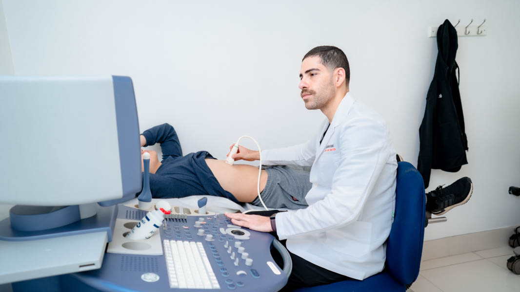
Ultrasound
Non-invasive real-time exploration of organs and tissues, widely used in obstetrics, cardiology, and abdominal studies.
- Dynamic and safe exploration
- Diagnostic versatility across multiple areas
- Ideal for obstetrics, abdomen, and soft tissue
- Immediate, accessible, and risk-free
- Color Doppler ultrasound, endocavitary ultrasound, biopsy-guided ultrasound, 3D ultrasound
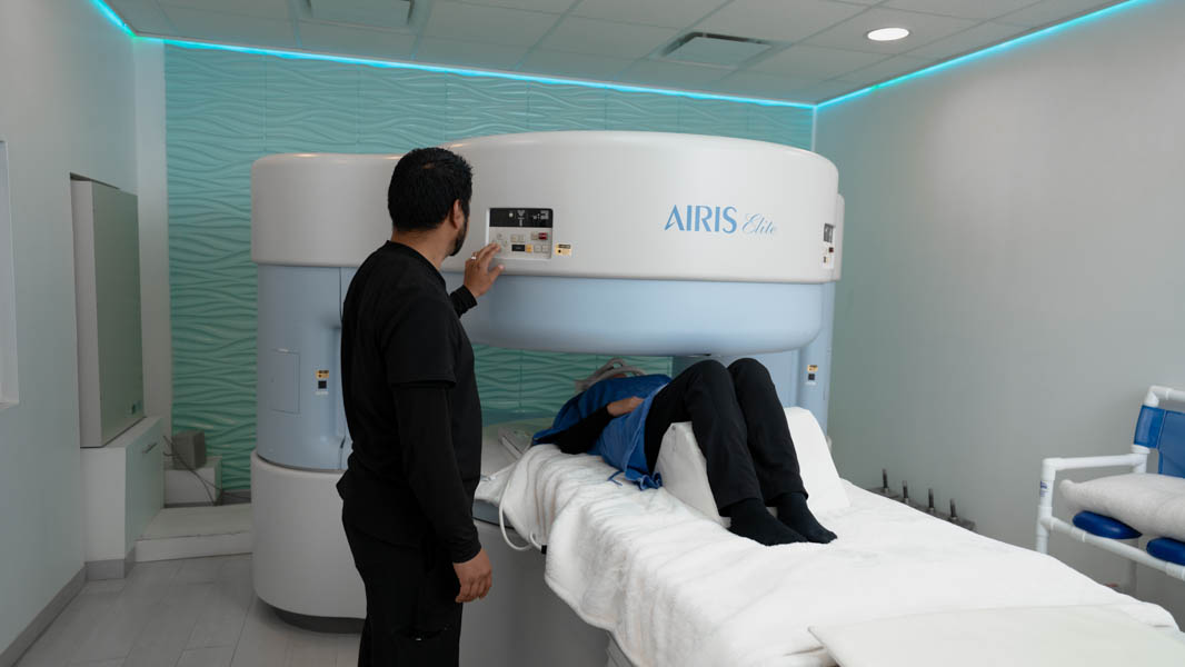
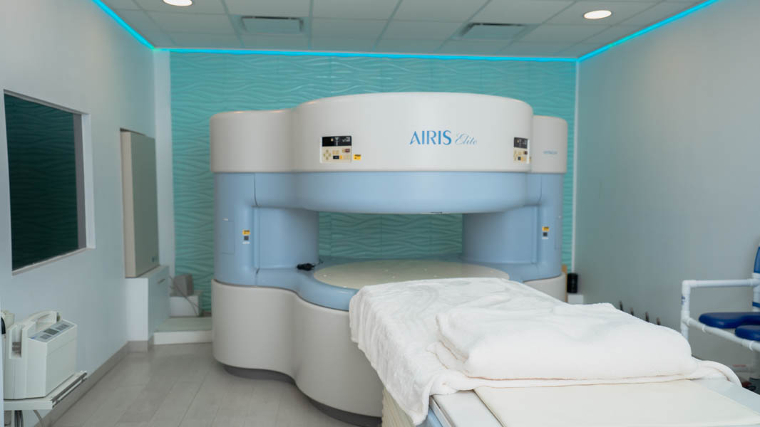
Magnetic Resonance Imaging (MRI)
High-resolution imaging of organs and tissues without radiation, providing key information for neurological, musculoskeletal, and cardiovascular diagnoses.
- Multiplanar imaging without radiation
- Ideal for the nervous system and soft tissues
- Quiet and precise scanning
- Advanced medical interpretation
Gastrography
A non-invasive study that allows visualization of the upper gastrointestinal tract through X-rays of the esophagus, stomach, and duodenum. It is used to detect structural alterations such as narrowing, masses, or hernias, as well as to evaluate symptoms like abdominal pain, reflux, or the presence of blood in the stool.
The patient ingests a contrast medium that highlights the internal walls of these organs, enabling real-time analysis through dynamic imaging.
- Clear visualization of the esophagus, stomach, and duodenum
- Non-invasive technique with ingested contrast
- Detection of masses, ulcers, and obstructions
- Useful evaluation for reflux, pain, or bleeding
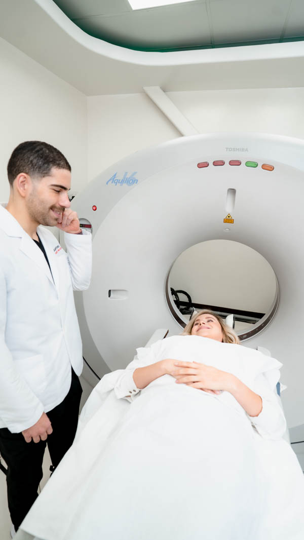
Computed Tomography (CT Scan)
Imaging study that combines multiple X-rays taken from different angles to generate cross-sectional views of the body. It allows for high-precision exploration of specific areas such as the brain, chest, spine, and abdomen. Its usefulness ranges from detecting fractures and internal bleeding to identifying tumors and various types of cancer.
It is a key tool in planning medical, surgical, or radiation treatments thanks to its ability to display internal structures with a high level of detail.
- Detailed exploration of brain, chest, spine, and abdomen
- Detection of fractures, tumors, cancer, and hemorrhages
- Precise imaging through cross-sectional slices
- Essential support in clinical treatment planning
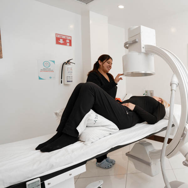
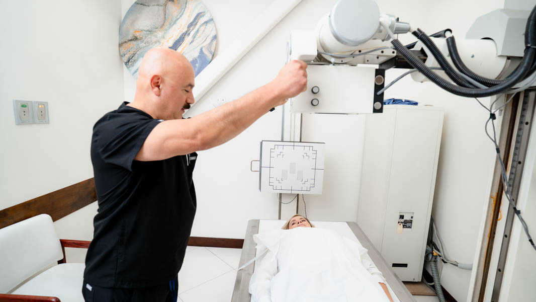
General X-Ray
Diagnostic study based on X-rays that allows the observation of internal body structures, such as bones and some organs. The procedure is quick, painless, and requires the patient to remain still for a few seconds to obtain clear images.
- Direct projection of bone tissues and specific areas
- Short and comfortable procedure
- High-definition results in minutes
- Interpretation validated by specialized radiologists
Home Radiology
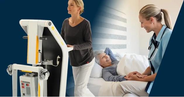
Imaging services in the comfort of your home, ideal for patients with limited mobility, ensuring accurate diagnoses without the need for transportation.
- Studies without the need for travel
- Care designed for reduced mobility
- High-performance portable equipment
- Digital results within a few hours
Digital Delivery of Studies with PACS
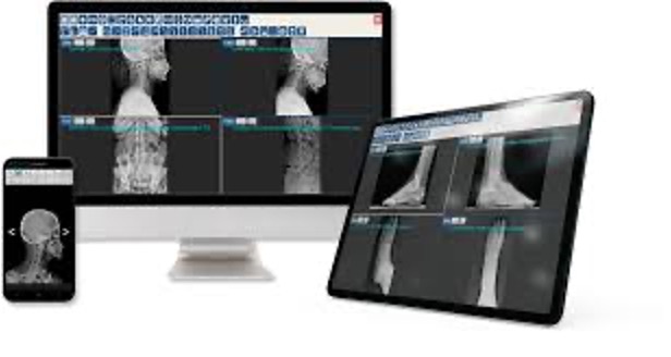
We use PACS, a system that allows your studies to be delivered in the cloud, digitally, securely, and interactively. This eliminates the need for film, facilitates remote access, and enhances your medical experience.
- Remote and immediate access to your studies from any device
- Digital and interactive viewing in high quality
- No film usage, more eco-friendly and efficient
